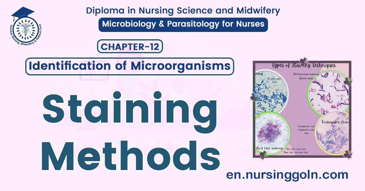
Staining Methods – Basic microbiology, parasitology, and immunology; nature, reproduction, growth, and transmission of common microorganisms and parasites in Bangladesh; prevention including universal precaution and immunization, control, sterilization, and disinfection; and specimen collections and examination. Students will have an understanding of common organisms and parasites caused human diseases and acquire knowledge about the prevention and control of those organisms.
Staining Methods
Definition of Staining Methods
Staining is an auxiliary technique used in microscopy to enhance contrast in the microscopic image. Stains and dyes are frequently used in biology and medicine to highlight structures in biological tissues for viewing, often with the aid of different microscopes.

Importance of Staining in Diagnostic Microbiology
- Identification of different bacterial species
- To know about the details morphology of the bacteria by different types of staining.
- To select proper antibiotics, as we know there are wide variation of antibiotic sensitivity between Gram positive, Gram negative & acid-fast bacteria. For example- Gram positive bbacteria are more sensitive to ẞ-lactam antibiotics than Gram-negative bacteria. So, identification of bacteria whether it is Gram positive or Gram negative is important prior to start freatment.
Classification of Staining Used in Microbiology
1. Simple stains: In this technique, a single staining substance is used.
Example:
- Methylene blue
- Crystal violet
- Basic fuchsine etc
2. Differential stains: Two stains with differing affinities to different bacteria are used in differential staining techniques.
Example:
- Gram stain.
- Ziehl- Neelsen’s stain / acid fast stain.
- Alberts stain – To see metachromatic granules of C. diphtheriae.
- Giemsa stain – To detect Chlamydia.
- Wayson’s stain to detect Yersinia pestis.
3. Negative stain:
Example:
- India ink stain for detection of capsule of Cryptococcus neofonnens fungus.
4. Fluorescent stains:
Example:
- Auramine and rhodamine to detect mycobacteria
5. Impregnation stain:
Example:
- Silver impregnation for Treponema pelidum
6. Special stains:
Example:
- Stain to see endospore
- Stain to see flagella
Principle of Gram Staining
Gram staining depends on the structures of bacterial cell wall & cytoplasmic membrane. Gram-positive bacteria have thick peptidoglycan layer on their cell wall, so they can resiste decolourization of primary stain by alcohol or acetone mixture, and is not stained by a counter stain. But Gram-negative bacteria have a thin peptidoglycan layer, so they cannot prevent decolourization by alcohol or acetone mixture, and so they are stained with a counter stain.

Steps of Gram staining
1. Preparation & fixation of smear: At first clean a slide and make a thin film with the 12.0 supplied specimen. Then dry it in the air. Fix the film by slowly passing the slide 3-4, wen times through a flame to song
2. Primary staining: Cover the film with gention or crystal violet and kept for 1 minute. It is then washed with tap water.
3. Mordanting: The slide is covered with a solution of Gram’s iodine / Lugol’s iodine for 1-
2 minutes.
4. Decolorizing: It is done by acetone or alcohol and is continued till the violet-color comes out from the smear. The slide then washed with tap water.
5. Counter staining: Cover the slide with dilute carbol fuchsin for 20-30 seconds. Wash with water and dry in air.
