The bones of the pelvic- The course is designed for the basic understanding of anatomical structures and physiological functions of human body, musculoskeletal system, digestive system, respiratory system; cardiovascular system; urinary system, endocrine system, reproductive system, nervous system, hematologic system, sensory organs, integumentary system, and immune system.
The aim of the course is to acquire knowledge and skills regarding anatomy and physiology.
The bones of the pelvic
The pelvic (hip) girdle consists of the two hip bones, also called coxal bones. The pelvic girdle provides a strong, stable support for the vertebral column, protects the pelvic viscera, and attaches the lower limbs to the axial skeleton. The hip bones are united to each other in front at a joint called the pubic symphysis posteriorly they unite with the sacrum at the sacroiliac joint.
Together with the sacrum and coccyx, the two hip bones of the pelvic girdle form a basinlike structure called the pelvis (plural is pelvises or pelves). In turn, the bony pelvis is divided into upper and lower portions by a boundary called the pelvicbrim.
False (greater) pelvis: The part of the pelvis above the pelvic brim is called the false (greater) pelvis. The false pelvis is actually part of the abdomen and does not contain any pelvic organs, except for the urinary bladder, when it is full, and the uterus during pregnancy.
True (lesser) pelvis: The part of the pelvis below the pelvic brim is called the true (lesser) pelvis. The true pelvis surrounds the pelvic cavity. The upper opening of the true pelvis is called the pelvic inlet, and the lower opening of the true pelvis is called the pelvic outlet.
The pelvicaxis is an imaginary curved line passing through the true pelvis; it joins the central points of the planes of the pelvic inlet and outlet. During childbirth, the pelvic axis is the course taken by the baby’s head as it descends through the pelvis.
(Ref-J. TORTORA, The essentials of anatomy and physiology, 8th edition, P-149.)
The pelvis (os coxae)
The pelvis is a combination of bones. It is comprised of the two os coxae, or coxal bones, joined posteriorly by the sacrum and anteriorly by a piece of fibrocartilage called the pubic symphysis. The pelvis forms unique landmarks that are not found on the pelvic girdles.
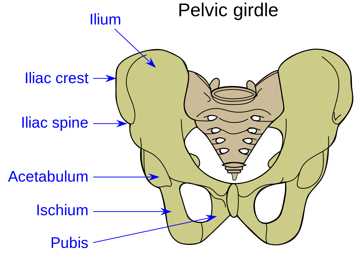
In the adult, the pelvis (os coxae) is formed by the fusion of three bones: ilium, ischium, and pubis Theunion of these three bones occurs at the acetabulum. The pairedos coxae articulate posteriorly with the sacrum and anteriorly with the pubic symphysis.
The following structures are formed within the fused os coxae.
- Acetabulum A cup-shaped socket into which the ballshaped head of the femur fits firmly.
- Obturator foramen, Covered by a flat sheet of connective tissue called the obturator membrane. A small opening located at the top of the membrane provides a route through which the obturator nerve, artery, and vein course.
- Greater sciatic notch. Located between the posterior inferior iliac spine and the ischial spine. The sacrospinous ligament converts the notch into the greater sciatic foramen, where the piriformis muscle, sciatic nerve, and pudendal neurovascular structures course.
- Lesser sciatic notch. Located between the ischial spine and the ischial tuberosity. The sacrotuberous ligament converts the notch into the lesser sciatic foramen.
- Pubic symphysis. Fibrocartilage connecting the two pubic bones in the anterior midline of the pelvis.
- Pelvic inlet. The superior aperture of the pelvis. The pelvic inlet is oval shaped and bounded by the ala of the sacrum, arcuate line, pubic bone, and symphysis pubis. The pelvic inlet is traversed by structures in the abdominal and pelvic cavities.
- Pelvic outlet. The inferior aperture of the pelvis. The pelvic outlet is a diamond-shaped opening formed by the pubic symphysis and sacrotuberous ligaments. Terminal parts of the vagina and the urinary and gastrointestinal tracts traverse the pelvic outlet. The perineum is inferior to the pelvic outlet. The pelvic bone is formed by the fusion of three bones: ilium, ischium, and pubis.
ILIUM
- Iliac crest. Thickened superior rim.
- Iliac fossa.Concave surface on the anteromedial surface.
- Anterior superior iliac spine. Anterior termination of the iliac crest. Serves as an attachment site for the sartorius and tensor fascia lata muscles.
- Anterior inferior iliac spine. Serves as an attachment site forthe rectus femoris muscle.
- Posterior superior iliac spine. Posterior termination of theiliac crest.
- Posterior inferior iliac spine. Forms the posterior border ofthe ala of the sacrum.
ISCHIUM
- Ischial tuberosity. A large protuberance on the inferior aspect of the ischium for attachment of the hamstring muscles and for supporting the body when sitting.
- Ischial spine. A pointed projection that separates the greater and lesser sciatic notches.
- Ischial ramus. A bony projection that joins with the inferior pubic ramus to form the ischiopubic ramus (conjoint ramus).
PUBIS
- Pubic tubercle. A rounded projection on the superior ramus of the pubis.
- Superior pubic ramus. A bony projection that forms a bridge from the acetabulum to the ischiopubic ramus, and thus the ischium. The crest on the superior aspect of the superior pubic ramus is the pectineal line, which serves as part of the border for the pelvic inlet and as an attachment site for muscles.
- Inferior pubic ramus. A bony projection that forms a bridge from the superior pubic ramus to the ischial ramus. The inferior pubic ramus serves as an attachment site for muscles of the lower limb
(Ref:- D. Morton, K. Albertine, The big picture Gross Anatomy, 2011, P-80)
Types of pelvis (pelvic shape)
There are four main types of pelvis, the prevalence dependent on sex and race. Forexample the relative frequencies in white females is:
- Gynaecoid round with enlarged transverse diameter – normal female type-41.4%
- Android-heart shaped in a woman may present hazards to normal delivery of a baby- 32.5%
- Anthropoid-long AP diameter -23.5%
- Platypelloid-long transverse diameter -2.6%
(Ref-C.W.F. Burnett FRCS, The Anatomy and Physiology of Obstetrics, 6 edition, P-81,82)
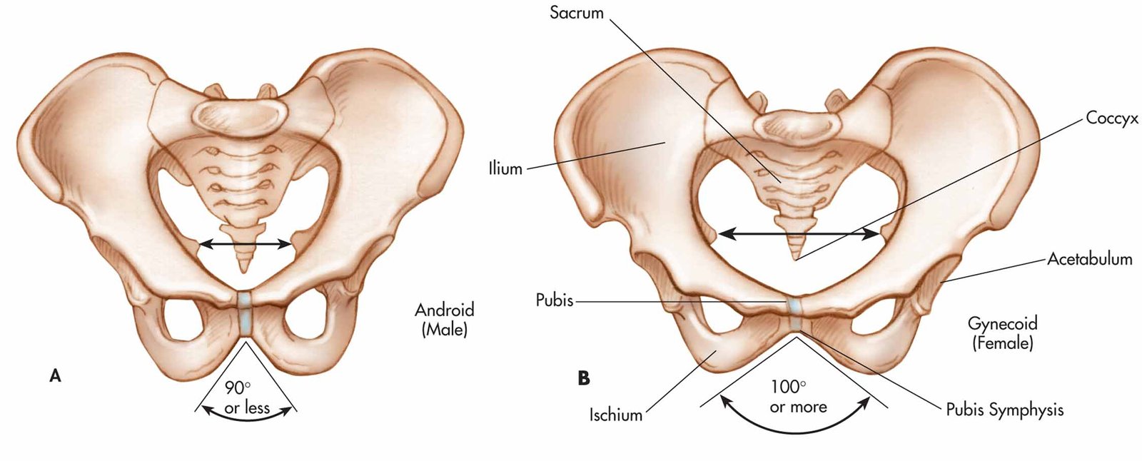
The bones of a male are generally larger and heavier than a female. The articular ends are thicker in relation to the shafts. In addition, because certain muscles of the male are larger than those of the female, the points of muscle attachment tuberosities, lines, and ridges are larger in the male skeleton. Many significant structural differences between the skeletons of females and males are related to pregnancy and childbirth.
Because the female’s pelvis is wider and shallower than the male’s, there is more space in the true pelvis of the female, especially in the pelvic inlet and pelvic outlet, which accommodate the passage of the infant’s head at birth. Several of the significant differences between the female and male pelvis are shown in below…..
(Ref-J. TORTORA. The essentials of anatomy and physiology, 8th edition, P-155)