Concept about Osteomyelitis – An orthopedic nurse is a nurse who specializes in treating patients with bone, limb, or musculoskeletal disorders. Nonetheless, because orthopedics and trauma typically follow one another, head injuries and infected wounds are frequently treated by orthopedic nurses.
Ensuring that patients receive the proper pre-and post-operative care following surgery is the responsibility of an orthopedic nurse. They play a critical role in the effort to return patients to baseline before admission. Early detection of complications following surgery, including sepsis, compartment syndrome, and site infections, falls under the purview of orthopedic nurses.
Concept about Osteomyelitis
Definition of Osteomyelitis:
Osteomyelitis is an inflammation of the bone, bone marrow, and surrounding soft tissue caused by the pyogenic (pus-producing) bacteria.
Or
Osteomyelitis is defined as a supportive process of the bone caused by pyogenic organisms or simply a pyogenic infection of the cancellous portion of the bone.
[Ref-John Ebnezar’s “Textbook of Orthopedics” 4 edition page-540]
Ог
Infection of all parts of the bone (Bone & bone marrow) is called osteomyelitis.
[Ref-Bailey & Love’s “Short Practice of Surgery” 25th edition page-546]
Or
Osteomyelitis is infection and inflammation of the bone or bone marrow.
[Reference: en.wikipedia.org]
Or
Osteomyelitis is an acute or chronic inflammatory process of the bone and its structures secondary to infection with pyogenic organisms.
Common joints affected in the osteomyolitis patients: In case of haematogenic spread the commonly affected joint are:
1. Hip joint.
2. Knee joint.
3. Ankle joint.
Mode of infection of the osteomyelitis patients: The mode of infection of osteomyolitis pts are:
1. Direct through the traumatic wound.
2. Haematogenous- from a infective focus from different parts of the bod, like skin infection.
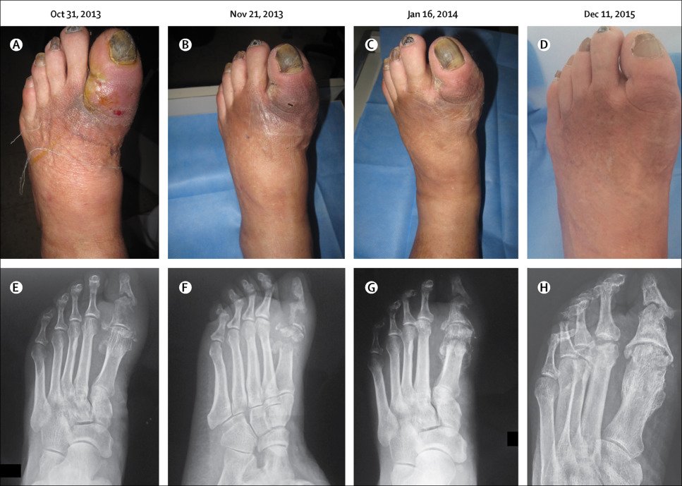
3. Contigious (from neibouring tissue). E.g: Diabetic foot ulcer.
4. Post traumatic osteomyolitis. E.g: open fracture.
Pathophysiology of osteomyelitis:
1. The infection results in the formation of abscess at the region of metaphysis.
2. The pus so finds its way out through the area of least resistance.
3. In children less than 2 years periosteum is loosely attached to the cortex and hence forms a potentially weak point.
4. The subperiosteal abscess so developed will either spread through the tissues or drain to the outside by forming a sinus breaking the skin or it will percolate down towards the diaphysis between the periosteum and the cortex and enter the shaft through the widened haversian pores due to anoxia.
5. The growth plate limits spread to the joint.
6. Between 2 and 16 years, periosteum is firmly attached to the cortex and with the growth plate still present; the pus has to spread towards the diaphysis at a slow pace.
7. Above 16 years, the growth plate has disappeared, the periosteum is fir adherent, and the pus spreads towards the diaphysis very slowly
Classification of osteomyelitis:
A. On the basis of duration/ clinical cause there are three types of osteomyelitis such as:
1. Acute osteomyelitis (<2 weeks): It is acute bacterial infection of the bone. When infection is carried by the blood, the onset is usually sudden occurring with a chill, high fever, rapid pulse and general malaise. E.g.
i. Haematogenous acute osteomyelitis: It is acute bacterial infection when carried by the
blood.
ii. Post traumatic acute osteomyelitis It occure due tocomplication from –
- Open fracture.
- Surgical wound.
- Penetrating injury.
2. Subacute osteomyelitis (2-3 weeks): Sub acute osteomyolitis is usually caused by staphylococcus aureus. The patient complains of pain without constitutional symptoms. Temperature may be increased or normal. It is not detected until at least two weeks has elapsed. E.g.:
✔Osteomyolitis with host resistance.
✔ Osteomyolitis with lowered bacterial resistance.
3. Chronic osteomyelitis ( 3 weeks) The patient with a chronic osteomyelitis, pres with continuously draining sinus or experiences recurrent periods of pa inflammation, Sub acute osteomyolitis is caused by staphylococcus aureus. The patient complains of pain without constitutional symptoms. Temperature may be increased or normal. It is not detected until at least two weeks has elapsed. E.g.:
✔Osteomyolitis with host resistance.
✔ Osteomyolitis with lowered bacterial resistance welling and drainage reduced blood supply. E.g.:
i. Specific: T.B. Syphilis, actinomycetes.
ii. Non-specific: Staphylococcus, streptococcus, salmonella, E. coli etc.
B. On the basis of aetiology:
a. Bacterial osteomyolitis (most common):
1. Pyogenic osteomyolitis.
2. Tubercular osteomyolitis.
3. Skeletal osteomyolitis.
b. virial osteomyolitis.
c. Fungal osteomyolitis.
d. Parasite osteomyolitis
C. On the basis of route of entry:
a) Haematogenous osteomyolitis: Infection is carried out by the blood.
b) Post traumatic osteomyolitis: E.g: open fracture.
Agent factors responsible for osteomyelitis: The ecological factors causing osteomyolitis.
These are:
A. Agent factors.
B. Host Factors:
C. Environment (General /local).
A. Agent Factors:
The following myriad of incriminating organisms is responsible for its causation:
1. “S” series organisms: “S” denotes severe osteomyelitis and those organisms causing it start with the letter “S”.
a) Staphylococcus aureus (60-85%): This is the most common organism causing acute osteomyelitis.
b) Streptococcus hemolyticus (8-10%).
c) Salmonella: Osteomyelitis is relatively rare and presents an interesting picture as most of its features start with “S”:
✔Several bones involved.
✔ Symmetrical involvement of bones.
✔Severe osteomyelitis.
✔Spine may be involved.
✔ Sickle cell anemia present.
✔Stool culture may be positive.
2. P-series organisms: Their mode of entry is through punctured wounds.
a. Pseudomonas.
b. Pneumococcus.
3. C-series: C denotes compound fractures.
a. Clostridium welchii.
b. Coliforms (E. coli).
4. B-series:
a. Brucella bacillus.
5. H-series:
a) Hemophilus influenzae (7 months to 4 years). This is known to cause osteomyelitis in the age group of
7 months to 4 years.
b) Osteomyelitis.
c) Traumatology & Orthopedic Nursing.
6. T-series:
a. Treponema pallidum (Syphilitic osteomyelitis).
b. Tubercle bacillus (Mycobacterium). Fungal osteomyelitis (ABC).
c. Actinomycosis.
d. Blastomycosis.
e. Cryptococcosis and coccidiodomycosis. These usually cause chronic osteomyelitis
B. Host Factors:
1. Age:
B. Host Factors:
a) Children: Incidence is 88% due to prone for injury and fall.
b) Adults: 12% is common in adult.
2. Sex: Male Female 4:1
3. Economic factors: Low socioeconomic group are more susceptible.
C. Environmental factors:
a) General factors: Anaemia Infection Poor nutrition Poor immune status
b) Local factors: Responsible for localization of infection at metaphysic especially in children. E.g.:
- Hair pin bend vessel.
- Metaphyseal haemorrhage.
- Defective phagocytosis.
- Anorexia.
- Rapid growth at metaphysic.
Predisposing factors for acute osteomyolitis:
a. Blood borne foci. (Boil septicaemia).
b. Age-Infancy & Childhood.
e. Sex-M:F-4:1.
d. Trauma-50% case.
e. Poor nutrition.
f. Unhygienic Surroundings.
Signs and symptoms of osteomyelitis:
A) History:
1. H/O of boils, minor injury, repeated infection.
2. H/O previous fractured 3-4 weeks.
3. H/O PTB, sickle cell diseases, dental, urinary, RTI.
4. Age: 5-15 years.
5. Sex-Male>Female.
B) Symptoms:
General Symptoms:
1. Fever (95%): this is the most common presenting symptom of osteomyolytris patient. The child usually has very high fever and is associated with profuse sweating, chills and rigors.
2. Sweating.: The patients presents with profuse sweating associated with fever.
3. Patient is usually in shock.
Local symptoms:
1. Local swelling (80%)
2. Swelling this usually follows the fever and may affects the end of the long bones. This swelling may acutely painful and the skin may appear red. Tenderness may present.
3. Limitation of movement (50%) limitation of movement: The child may not move the joint near the affected bone due to pain and swelling. In fact, the child may lie still without moving the joint and this is sometimes called a state of pseudoparalysis.
Clinical features:
A. Genera signs:
1. Increased temperature.
2. Increase pulse rate.
3. General feature of anaemia.
4. Signs of dehydration and shock.
5. Shock and toxicity may be present.
B. Local signs:
1. Tenderness (80%).
2. Local erythema (50%).
3. Raised temperature (50%).
4. Fluctuation present (20%).
5. Effusion (10%).
6. Decreased movements (50%.
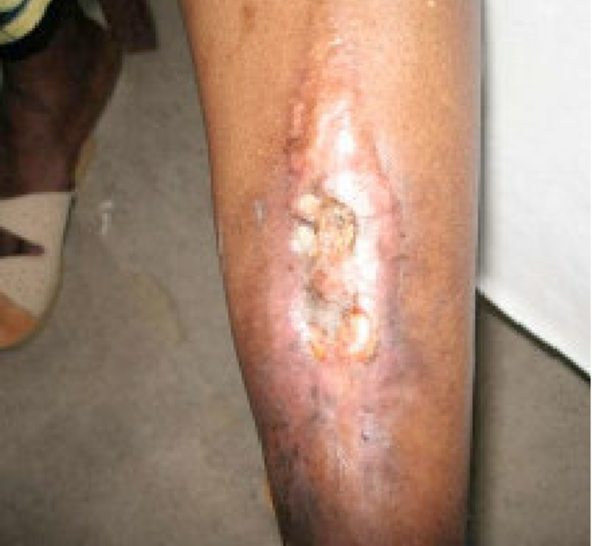
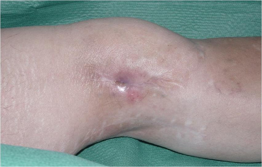
7. Draining around the sinus at open fracture /end of long bone. (in caese of chronic osteomyolitis)
8. X-ray: Thickening of the bone.
9. Affected site: femur, tibia, humerus(at the metaphysis)
10. Fluctuation test may positive.
[Ref-John Ebnezar’s “Textbook of Orthopedics” edition page]
Chronic osteomyelitis:
Any osteomyelitis lasting for more than three weeks is termed as chronic. Chronic osteomyelitis can arise from any one of the following ways:
1. Sequelae of acute osteomyelitis (5-10%).
2. Following compound fractures.
3. Following surgery on bones and joints.
4. Chronic from the beginning (e.g. tuberculosis, syphilis, Brodie’s abscess).
5. Anaerobic organisms (sclerosing osteomyelitis of Garre).
6. Fungal osteomyelitis.
[Ref-John Ebnezar’s “Textbook of Orthopedics” 4 edition page-546]
Characteristics of acute osteomvelitis:
1. Disease is common in children.
2. Staphylococcus aureus is the common organism.
3. Metaphysis is involved.
4. Fever is the common presenting system.
5. Bone scan helps in early diagnosis.
6. Conservative management is the mainstay of treatment. In addition, ninety percent resolve
[Ref-John Ebnezar’s “Textbook of Orthopedics” edition page->546]
Differential diagnoses of osteomvelitis:
1. Acute septic arthritis:
d) Tenderness mainly in the joint.
e) Joint is swollen
f) joint movements are severely restricted and more painful in acute septic arthritis.
2. Scurvy:
a) Features of pseudoparalysis,
b) bleeding gums,
c) tender limbs, etc are the features.
3. Haemarthrosis:
a) Usually haemophylia.
b) H/O bleeding.
H/O bleeding during tooth extraction.
4. Acute anterior poliomyelitis:
Here pain and tenderness are spread throughout the muscle mass, whereas in osteomyelitis tenderness is greatest on direct pressure over the bone.
5. Cellulitis:
It is difficult to differentiate from acute osteomyelitis; however, cellulites has no edge, no fluctuation, no pus and no limits.
6. Other differential diagnosis:
✔ Erysipelas.
✔Erythema nodosum.
✔ Ewing’s sarcoma.
✔Sickle cell anemia.
[Ref-John Ebnezar’s “Textbook of Orthopedics” edition page-545]
Acute osteomyelitis is common in children :
Acute osteomyelitis is common in children because
1. Inadequate immunity, so trauma in malnourished children causes colonization of bacteria.
2. All the bones are rapid growing & more vascular.
3. Thin, more elastic periosteum in children.
Source of infection in acute haematogenous osteomyelitis:
1. URTI: e.g. tonsillitis, adenitis.
2. Boils and abscess.
3. Bacteraemia, septicemia or pyemia
4. Bones commonly affected are: Femur, Tibia, Humerus.
Complications of acute osteomyelitis:
1. Local:
i. Chronic osteomyelitis.
ii. Pyogenic arthritis,
iii. Pathological fracture,
iv. Deformity.
V. Retardation of growth arrest.
2. General:
i. Septicaemia,
ii. Pyaemia.
Complications of chronic osteomyelitis:
1. Pathological fracture.
2. Persistent discharge of pus.
3. Septicaemia.
4. Septic arthritis.
5. Amyloidosis.
6. Development of sequamous cell carcinoma in a sinus (Marjolin’s ulcer).
7. Acute flare up of inflammation.
[Ref-Adams/11/78]
Treatment of active acute pvogenic osteomyelitis:
A) General management:
1. Absolute bed rest.
2. Reassurance to the patient and his/her family members.
3. Protect the affected part with splints to alleviate pain and spasm.
4. Take care of the splient side.
5. Tapid sponging.
4. Symptomatic treatment will be given according to doctor’s advice.
5. Intravenous fluid may be given.
6. Cast may be apply for prevent fracture of weakened bone.
7. Dressing will be change frequently.
8. Apparatus of external fixation will be check frequently.
9. Maintain muscle and joint strength.
10. Care of the limb and regular physiotherapy and exercise. 11. Adequate vitamin and nutrient containing diet will be given.
12. Advice to intake blood building food and frequent small meal.
13. Personal hygiene must be maintained.
14. Sign and symptoms will be observe and record on a chart.
15. Encourage to the patient and his/her family members for surgical treatment.
16. All investigations will be done and patient prepare for operation when failure conservative treatment.
17. Cast may be apply for prevent fracture of weakened bone.
18. Dressing will be change frequently.
19. Apparatus of external fixation will be check frequently
20. Maintain muscle and joun srengur.
21. Care of the limb and regular physiotherapy and exercise. 22. Psychotherapy and counseling to the patient.
23. Pre-operative medication and care will be given.
24. Post operative care will be given.
25. Health education will be given to the patient and his/her family members.
A) Medical management:
ii. Splint is applied
ii.Antibiotic therapy: Antibiotic according to C/S report:
a) Cloxacillin 500mg 6 hourly (Staphylococcus aureus)
b) Ampicillin 250mg 6 hourly(H. influenza)
e) Arythromycin
d) Cefotaxime
e) Cefadroxyl
f) Ciprofloxacin
g) cephalosporin
iii. fluid to correct shock and hypovolaemia.
iv. Blood transfution if needed.
v. Analgesics.
B) Surgical management:
a) Aspiration: Collection of pus from the site of the infection for c/s and proper antibiotic therapy.
b) Incision and drainage: An incision is made over the tender area and carried down to the bone where pus is usually found to the deep periosteum. Draining the abscess.
c) Multiple drill hole: If the abscess is sub-periosteal, multiple drill holes in the cortex to drain the pus.
d) Small bone window: If multiple drill hole do not drain the pus,a small window of bone is carried out from
e) cortex for pus evacuation. In case chronic tubercular osteomyolitis:
i. Sequestrectomy. (Removal of sequestrum)
ii. Saucerization & filling the cavity by muscle or soft tissue, bone grafting.
iii. Surgical evacuation & curettage of the cavity
iv. Packing with cancellous bone clips(for moderate size cavity)
Nursing care for an osteomyelitis patient:
1) Nursing assessment.
2) Nursing diagnosis:
a) Impaired physical mobility related to bony involvement.
b) Acute pain related to presence of abscess in the bone and soft tissue.
c) Risk of infection related to pus formation.
d) Spread of infection related to contact with other area of the body.
e) Impaired skin integrity related to incision and surgery.
f) Anxiety related to economic cost of prolong treatment.
3) Nursing intervention:
a) Promoting physical mobility by:
1) Rest the affected part.
2) Give splint for immobilization.
3) Regular changing of the splint or cast.
4) Care of splint.
5) Advise the patient his her finger physiotherapy.
6) Avoid excessive activity.
7) Avoid pressure on the affected area.
b) Relieving pain:
1) Restrict movement on the affected part.
2) Splint is use full to reduce the pain.
3) Elevation of the affected part to reduce swelling as well as pain reduction
4) Give analgesics according to doctors order.
c) Prevention of infection:
1) Regular observation of the affected area.
2) Regular dressing with antiseptic solution.
3) Provide proper antibiotic to prevent further complication.
4) Aspiration of pus.
5) Incision and drainage of pus from the abscess cavity.
6) Strict asceptic technique should be used.
d) Prevention of spread of infection:
1) Proper dressing the area with antiseptic solution.
2) Proper hand washing.
3) Always remember about handling of the pus.
4) Never contact the pus with other body surface.
5) Personal hygiene must be maintained.
e) Reduction of anxiety:
1) Reassurance the patient.
2) Psychotherapy.
3) Tell him about modern treatment method and way of prognosis.
4) Proper counseling to the patient.
f) Maintain proper nutritional status.
g) Operative management:
1) Preoperative management:
i. Clean and shave the operative area.
ii. Clean the operative area with hot water and shop.
iii. Complete all the investigation.
iv. Check the G/A fitness.
v. Take the written consent.
vi. Adequate fluid must be given.
vii. Carefull about asthma, HTN, and DM.
viii. Open an I/V channel.
ix. Transfer the patient to the operation theater in proper time.
2) Operative management:
1. Check the name address, age site, of operation.
ii. Prepare the operation tolly.
iii. Use asceptic gloves,gown, and instrument. iv. Observe the patient during operation.
V. After complete the operation, transfer the patient to post operative ward.
3) Post operative management:
1. Proper positioning of the patient.
ii. Elevated the area of the operation.
iii. Assess pulse, respiration, blood pressure and temperature.
iv. Maintain intake and output chart.
v. Apparatus of external fixation check regularly.
vi. Proper wound dressing.
2 possible D/D:
1. Acute osteomyelitis.
2. Septic arthritis.
2 important investigations:
1. X-ray of right knee joint.
2. Joint aspiration & C/S.
BRODIE’S ABSCESS:
BRODIE’S ABSCESS is a localized form of chronic osteomyolitis, involves metaphysis and epiphyseal area, and common in young adult.
Baker’s cyst:
Baker ‘s cyst is simply a herniation of the synovial cavity of the knee, with the formation of fluid- filled sac extending backwards and downwards. It is not a primary condition but is always secondary to a disorder of the knee with persistent synovial effusion, such as rheumatoid arthritis or osteoarthritis. In long standing cases hernial sac is much elongated, and may extend a considerable distance down the calf. Clinically there a soft cystic bulge nears the midline behind the knee or in the upper calf. The underlying abnormality of the knee. With synovial effusion, will usually be obvious.
Treatment:
In most cases treatment should be directed towards the underling condition of the knee rather than to the cyst itself. Nevertheless if the cyst is extensive it is sometimes advisable to excise it
Diagnosis:
Acute pyogenic osteomyolitis. 2D/D:
1) Abscess in the thigh.
2) Ewing s tumor.
- Investigation: Please see above.
- Complication: Please see above.