Concept About Club Foot – An orthopedic nurse is a nurse who specializes in treating patients with bone, limb, or musculoskeletal disorders. Nonetheless, because orthopedics and trauma typically follow one another, head injuries and infected wounds are frequently treated by orthopedic nurses.
Ensuring that patients receive the proper pre-and post-operative care following surgery is the responsibility of an orthopedic nurse. They play a critical role in the effort to return patients to baseline before admission. Early detection of complications following surgery, including sepsis, compartment syndrome, and site infections, falls under the purview of orthopedic nurses.
Concept About Club Foot
Definition of club foot or Congenital talepes Equinovarus:
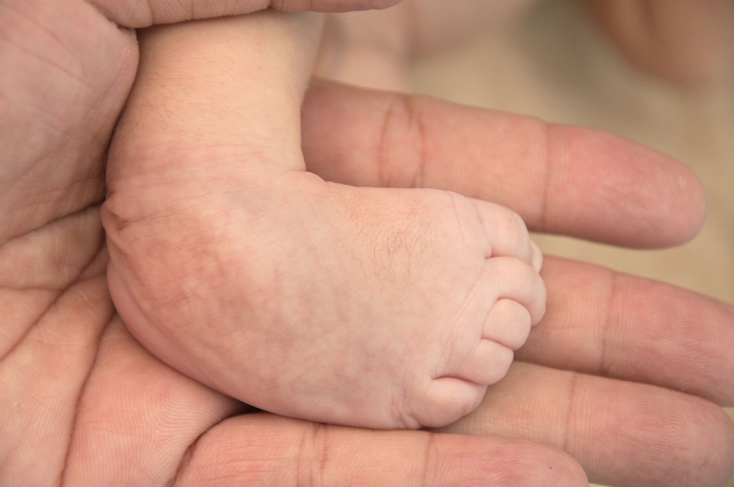
Club foot means, club like appeartance. Talepes comes from two Latin word: “Talus’ means Ankle and Pes means Foot. Original meaning: A deformity that causes the patient to walk on ankle.
[Ref-John Ebnezar’s, “Textbook of Orthopedics”, 4″ edition, P-503]
Or
It is a congenital deformity characterized by plantar flexin (equinus) and inversion (varus) of the ankle so that dorsiflexion of the foot is limited; and unlike normal babies, the dorsum of the foot can not be made to touch the front of shin.
[Ref- Mr Khan ‘s Essential of pediatrics, “, 4” edition, P-12]
Or
Club foot (Talepes equinovarus) is a combination of abnormalities of bony architecture and soft tissue. Fore foot adduction and supination, heel varus, ankle equines and medial deviation of the entire foot are 4 abnormal features presented with club foot.
Types of Congenital Talepes Equinovarus or club foot:
1. Osseous type: Clubfoot is associated with absence of tibia and fibula.
2. Muscular type: Arthrogryposis multiplex congenital contractures.
3. Neuropathic type: Due to spina bifida, etc
4. Idiopathic type: No apparent cause commonest variety.
[Ref-John Ebnezar’s, “Textbook of Orthopedics”, 4″ edition, P-503]
Types of foot deformities:
There are various types of foot deformities. These are given below:
1. Varus.
2. Equinovarus.
3. Calcaneovarus.
4. Equinus.
5. Calcaneus.
6. Cavus.
7. Valgus.
8. Calcaneovalgus.
9. Equinovalgus.
[Ref-John Ebnezar’s, “Textbook of Orthopedics”, 4″ edition, P-503]
Cause’s clubfoot:
In some cases, clubfoot is just the result of the position of the baby while it is developing in the mother’s womb (postural clubfoot). But more often clubfoot is caused by a combination of genetic and environmental factors that is not well understood. If someone in your family has clubfoot, then it is more likely to occur in your infant. If your family has one child with clubfoot, the chances of a second infant having the condition increase.
Clubfoot present at birth can point to further health problems because clubfoot can be linked with other conditions such as spina bifida. For this reason, as soon as clubfoot is noticed, it’s important that the infant be screened for other health conditions. Clubfoot can also be the result of problems that affect the nerve, muscle, and bone systems, such as stroke or brain injury.
Symptoms of clubfoot:
Clubfoot is painless in a baby, but it can eventually cause discomfort and become a noticeable disability. Left untreated, clubfoot does not straighten itself out. The foot will remain twisted out of shape, and the affected leg may be shorter and smaller than the other. These symptoms become more obvious and more of a problem as the child grows. There are also problems with fitting shoes and participating in normal play. Treatment that begins shortly after birth can help overcome these problems. Diagnosis:
Ultrasound done while a baby is in the womb can sometimes detect clubfoot. It is more common for your doctor to diagnose the condition after the infant is born, though, based on the appearance and mobility of the feet and legs. In some cases, especially if the clubfoot is due just to the position of the growing baby (postural clubfoot), the foot is flexible and can be moved into a normal or nearly normal position after the baby is born.
In other cases, the foot is more rigid or stiff, and the muscles at the back of the calf are very tight. X-rays may not be helpful to confirm the diagnosis. Some of the baby’s foot and ankle bones are not fully ossified (filled in with bony material) and do not show well on X-ray.
Aim of treatment:
1. Early correction.
2. Full correction.
3. Maintenance of correction and prevention of recurrence.
Treatment of club foot:
Conservative treatment:
a. Treat at birth Passive stretching done by mother with each feed. Varus correction followed by correction of equinus deformity.
b. Plaster at thin soft skin not done until 7-10 days after birth.
c. After 7-10 days: Serial plaster.
d. Serial plaster can be tried till 6 month age.
e. Serial plaster done weekly intervals – 6 weeks.
f. Then biweekly – next 6 weeks or more.
g. After correction: Corrective shoe or splints for 1-2 years.
h. More than half of the cases corrected by only early serial plaster. i. Correction: Adduction 1, then heel varus then equinus.
Surgical management:
1. Soft tissue release:
a. Postero-medial release + Elongation of Tendoachilia. Effective upto4th year.
b. After 4 year: Postero-medial release (PMR) -if Dwyer’s lateral closedosteotomy.
c. Evan’s Calcaneocuboid decancellation or fusion
2. In case of older children or adolescents:
a. Talectomy.
b. Tendon transfer (T.A laterally),
c. Triple arthrodesis (After 12 years),
d. Treat by external fixators.
Nursing Management of club foot:
1. Management should be started as early as possible with the standard foot wear, may be in the first week of life.
2. Give mental support and reassure to the parents and tell them that, this disease is completely preventable.
3. Initial non-operative management is done by serial manipulation followed by immobilization in a plaster cast or adhesive tape or strapping or splinting.
4. Parents should be taught with adequate explanation and demonstration about the manipulation that can be done at home.
5. This initial management may be continued for two to six months of age.
6. Then assessment is necessary about the progress and to plan for further management.
7. Explanation to be given to the parents about details of surgical correction of the birth defect which usually performed at 4 to 9 months of age with postoperative immobilization before the child begins to walk.
8. Maintain nutritional status of the child – give exclusive breast feeding till 6 months.
9. Show acceptance of the infant, maintain composure and do not show negative emotion when handling the infant.
10. Encourage parental involvement in infants care: frequent holding, cuddling and playing.
11. Observe and assess reciprocity of cues between infant and parents to identify behavior that may need strengthening.
12. Discuss with parents about correct preparation formula.
13. Instruct newborn care to the parents.
14. Emphasize to follow up as appointment.
Club foot complex:
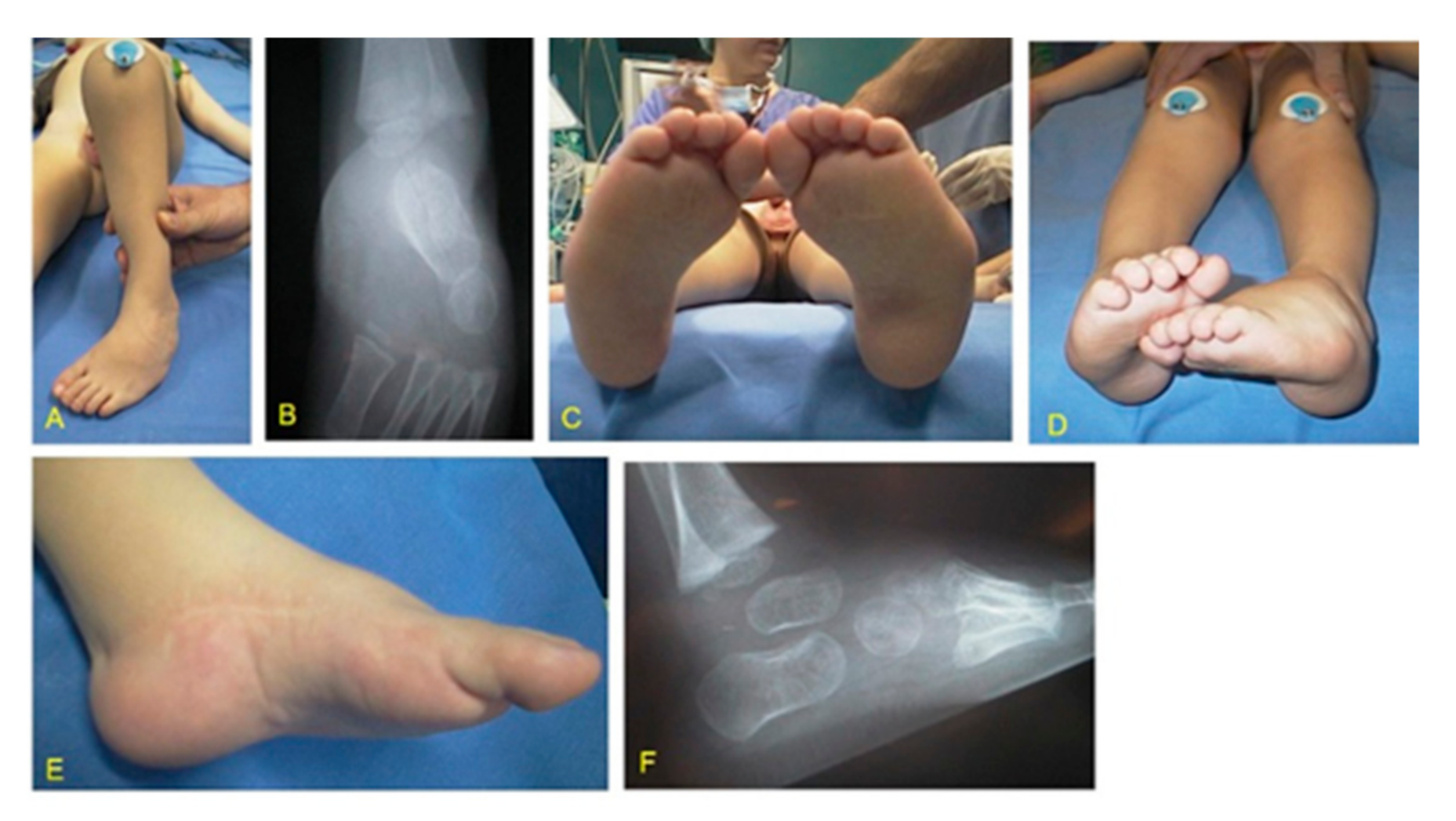
Congenital talipes equinovarus is a grotesque looking deformity of the foot. Five classical primary deformities are seen and in response to this, secondary deformities develop. The primary and secondary deformities together form the club foot complex.
Flow chart of club foot complex:
Club foot complex
↓
Secondary deformities
↓
1. Foot size is decreased to 50%
2. Medical border is concave, lateral border is convex.
3. Forefoot is plantarflexed upon hindfoot.
4. Skin is stretched over the dorsum of the foot.
5. Callosities are present over the dorsum of the foot.
6. Stumbling gait
7. Hypotrophic anterior tibial artery
8. Atrophy of muscles in anterioror posterior compartments of the leg.
↓
Primary deformities
1. Equinus
2. Varus
3. Cavus
4. Forefoot adduction
5. Internal tibial torsion
↓
Late changes
↓
1. Degeneration of joints
2. Fusion of joints
[Ref-John Ebnezar’s, “Textbook of Orthopedics”, 4″ edition, P-504]
Difference between CTEV and ATEV:
| Congenital Talepes Equinovarus | Acquired Talepes Equinovarus |
| 1. Present since birth. | 1. Not present from birth. |
| 2. May be associated with spina bifida. | 2. May be due to polio, cerebral palsy etc. |
| 3. Bilateral. | 3. Usually unilateral. |
| 4. Skin, subcutaneous tissue, muscles are normal. | 4. Tropic changes in the skin, muscles are flaccid or spastic. |
| 5. Transverse crease is seen across the sole on the medial side | 5. No transverse crease. |
| 6. Bones are normal in thickness. | 6. Bones are thinner than norma |
Indications of surgical management:
1. Inadequate response to serial plaster.
2. Rigid club foot.
3. Relapsed club foot.
4. Recurrent club foot (Associated with muscle imbalance).
5. Resistant club foot (Totally resistant).