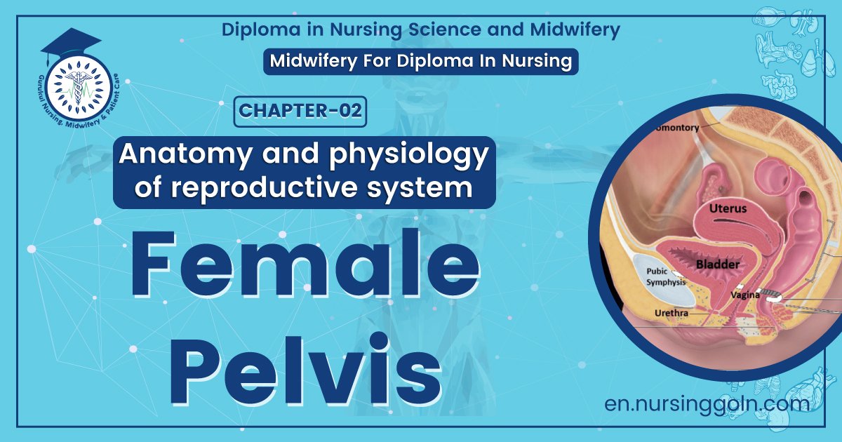
Female pelvis – This course is designed to understand the care of pregnant women and newborn: antenatal, intra-natal and postnatal; breast feeding, family planning, newborn care and ethical issues, The aim of the course is to acquire knowledge and develop competencies regarding midwifery, complicated labour and newborn care including family planning.
Female pelvis
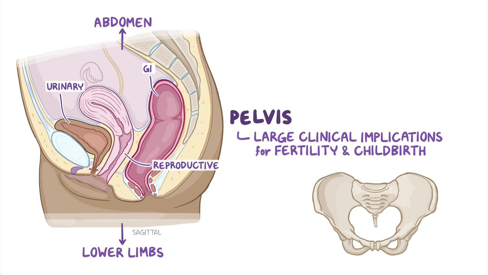
Figure: female pelvis
The pelvis is the lower part of the torso. It’s located between the abdomen and the legs. This area provides support for the intestines and also contains the bladder and reproductive organs. There are some structural differences between the female and the male pelvis. Most of these differences involve providing enough space for a baby to develop and pass through the birth canal of the female pelvis. As a result, the female pelvis is generally broader and wider than the male pelvis.
Female pelvis anatomy and function
Female pelvis bones:
Hip bones:
There are two hip bones, one on the left side of the body and the other on the right. Together, they form the part of the pelvis called the pelvic girdle.
The hip bones join to the upper part of the skeleton through attachment at the sacrum. Each hip bone is made of three smaller bones that fuse together during adolescence
- Ilium. The largest part of the hip bone, the ilium, is broad and fan-shaped. You can feel the arches of these bones when you put your hands on your hips
- Pubis. The pubis bone of each hip bone connects to the other at a joint called the pubis symphysis
- Ischium. When you sit down, most of your body weight falls on these bones. This is why they’re sometimes called sit bones.
The ilium, pubis, and ischium of each hip bone come together to form the acetabulum, where the head of the thigh bone (femur) attaches.
Sacrum
The sacrum is connected to the lower part of the vertebrae. It’s actually made up of five vertebrae that have fused together. The sacrum is quite thick and helps to support body weight.
Соссух
The coccyx is sometimes called the tailbone. It’s connected to the bottom of the sacrum supported by several ligaments.
The coccyx is made up of four vertebrae that have fused into a triangle-like shape.
Pelvic joints:
- Sacroiliac joints x 2
- Symphysis pubis
- Sacro-coccygeal joint
Features of the pelvis:
- Iliac crests
- Sciatic notch
- Obturator foramen
- Ischial tuberosities
- Ischial spines Pubic arch
- Superior and inferior pubic rami
Ref-DMW, Lesson plan volume 1/ page 28-29]
Features of the boundary of the brim:
- Promontory of sacrum
- Wings or alae of the sacrum
- Right and left sacroiliac joint
- Right and left iliopectineal line
- Right and left iliopectineal eminences
- Upper inner borders of left and right superior pubic rami
- Upper inner borders of pubic bodies
- Upper inner border of symphvsis pubis
Female pelvis muscles
Levator ani muscles
- The levator ani muscles are the largest group of muscles in the pelvis. They have several functions, including helping to support the pelvic organs.
- The levator ani muscles consist of three separate muscles:
- Puborectalis. This muscle is responsible for holding in urine and feces. It relaxes when you urinate or have a bowel movement.
- Pubococcygeus. This muscle makes up most of the levator ani muscles. It originates at the pubis bone and connects to the coccyx.
- Iliococcygeus. The iliococcygeus has thinner fibers and serves to lift the pelvic floor as well as the anal canal.
Coccygeus
This small pelvic floor muscle originates at the ischium and connects to the sacrum and coccyx.
[Ref-DMW, Lesson plan volume 1/ page 28-29]
Female pelvis organs
Uterus
The uterus is a thick-walled, hollow organ where a baby develops during pregnancy.ab
During the reproductive years, the lining of the uterus sheds every month during menstruation if you don’t become pregnant.
Ovaries
There are two ovaries located on either side of the uterus. The ovaries produce eggs and also release hormones, such estrogen and progesterone.
Fallopian tubes
The fallopian tubes connect each ovary to the uterus. Specialized cells in the fallopian tubes use hair-like structures called cilia to help direct eggs from the ovaries toward the uterus
Cervixe
The cervix connects the uterus to the vagina. It’s able to widen, allowing sperm to pass into the uterus.
In addition, thick mucus produced in the cervix can help to prevent bacteria from reaching the uterus.
Vagina
The vagina connects the cervix to the exterior female genitalia. It’s also called the birth canal, as the baby passes through the vagina during delivery.
Rectum
The rectum is the lowest part of the large intestine. Feces collects here until exiting through the anus.
Bladder
The bladder is the organ that collects and stores urine until it’s released. Urine reaches the bladder through tubes called ureters that connect to the kidneys.
Urethra
The urethra is the tube that urine travels through to exit the body from the bladder. The female urethra is much shorter than the male urethra.
[Ref-DMW, Lesson plan volume 1/ page 28-29]
Female pelvis ligaments:
Broad ligament
The broad ligament supports the uterus, fallopian tubes, and ovaries. It extends to both sides of the pelvic wall.
The broad ligament can be further divided into three components that are linked to different parts of the female reproductive organs:
- mesometrium, which supports the uterus
- mesovarium, which supports the ovaries
- mesosalpinx, which supports the fallopian tubes
Uterine ligaments
Uterine ligaments provide additional support for the uterus. Some of the main uterine ligaments include:
- the round ligament
- cardinal ligaments
- pubocervical ligaments
- uterosacral ligaments
Ovarian ligaments
The ovarian ligaments support the ovaries. There are two main ovarian ligaments:
- the ovarian ligament
- the suspensory ligament of the ovary
Pelvic brim dimensions:
- Anteroposterior
- Right and left oblique
- Transverse
Pelvic cavity dimensions:
- Anteroposterior
- Right and left oblique
- Transverse
Pelvic outlet dimensions:
- Anteroposterior
- Right and left oblique
- Transverse
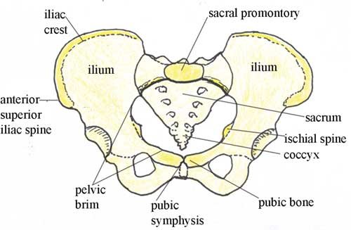
Figure: Female pelvis
Female pelvis conditions:
The pelvis contains a large number of organs, bones, muscles, and ligaments, so many conditions can affect the entire pelvis or parts within it.
Some conditions that can at can affect the female pelvis as a whole include:
- Pelvic inflammatory disease (PID). PID is an infection that occurs in the female reproductive system. While it’s often caused by a sexually transmitted infection, other infections can also cause PID. Untreated PID can lead to complications, such as infertility or ectopic pregnancy.
- Pelvic organ prolapse. Pelvic organ prolapse occurs when the muscles in the pelvis can no longer support its organs, such as the bladder, uterus, or rectum. This can cause one or more of these organs to press down on the vagina. In some cases, this can cause a bulge to form outside of the vagina.
- Endometriosis. Endometriosis occurs when the tissue that lines the inside walls of the uterus (endometrium) begins to grow outside of of the uterus. The ovaries, fallopian tubes, and other tissues in the pelvis are typically affected by the condition. Endometriosis can lead to complications, including infertility or ovarian cancer.
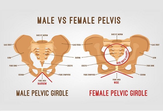
Distinct Features of Male and female Pelvis
Distinct Features of The Male Pelvis
- Male pelvis is smaller and narrower with heavier and thicker bones.
- It has a longer and narrower sacrum.
- The pelvis has a heart-shaped pelvic inlet.
- It consist of an acetabulum that is larger
- Ilium of male pelvis is more vertical with more curved iliac crest
- In Male pelvis, ischial tuberosity is longer, close together and more laterally projecting.
- The pelvis has a v-shaped pubic arch.
- It pelvis has a narrower sciatic notch.
- The coccyx of a male pelvis is projected inwards (less curved anteriorly) and immovable.
- Male pelvis is designed to support a heavy body with a stronger muscle structure.
- In male pelvis, the obturator foramen is round.
- A male pelvic bone is heavier, taller and much thicker
- The pelvic outlet in male pelvis is narrower.
Distinct Features of The Female Pelvis
- The bones are more delicate-thin and light
- The pelvis is less massive and more shallow
- The ilia are less sloped
- The superior aperture of the lesser pelvis (pelvic inlet) is larger, more nearly circular and has greater obliquity.
- The anterior iliac spines are more widely separated- thus the greater prominence of the hips laterally
- The cavity of the pelvis is shallow and wider.
- The obturator foramina are triangular I in shape and smaller, in size than the male circular foramina
- The acetabula are smaller and look more distinctly foward.
- The sciatic notches are wider and shallower.
- The pubic symphysis is is less deep
- The superior pubic ramus is longer than the width of the acetabulum
- Sacrum is shorter, wider and the upper part is less curved, so the sa sacral promontory is less imposing into the pelvic cavity
- The inferior aperture of the lesser pelvis (pelvic outlet) is larger and the coccyx is more moveable.
- The pubic arch is wider and more rounded than in male where it is an angle rather than an arch.
- The muscle attachments are more poorly marked.
- The spines of the ischia project less inward hence not protruding as much into the pelvic cavity.
- The Ischia tuberosities and the acetabula are more wider apart.
| Elements of Comparison | Male Pelvis | Female Pelvis |
| Size | Male pelvis is smaller and narrower with heavier and thicker bones. | Female pelvis is bigger and wide with lighter and denser bones. |
| Sacrum | Has a longer and narrower sacrum | Has a sacrum that is wider, shorter and less curved. |
| Pelvic Inlet/Brim | Has a heart-shaped pelvic inlet | Has pelvic inlet that is slightly oval in shape. |
| Acetabulum | The acetabulum is larger | The acetabulum is smaller |
| Ilium | Ilium of male pelvis is more vertical with more curved iliac crest. | The Ilium of female pelvis is less vertical with less curved iliac crest. |
| Ischial tuberosity | The ischial tuberosity is longer, close together and more laterally projecting. | The ischial tuberosity is shorter, farther apart and more medially projecting. |
| Pubic Arch | Has a v-shaped pubic arch | Has a pubic arch that is wider. |
| Sciatic Notch | Sciatic notch is narrower | Sciatic notch is wider |
| coccyх | Coccyx of a male pelvis is projected inwards (less curved anteriorly) and immovable. | Female pelvis has a flexible and straighter coccyх. |
| Purpose of Design | Male pelvis is designed to support a heavy body with a stronger muscle structure. | The female pelvis has been designed for purposes of childbearing and easier delivery. |
| Obturator foramen | The obturator foramen is round | The obturator foramen is oval. |
| Pelvic Bone | Male pelvic bone is heavier, taller and much thicker | Female pelvic bone is thinner and denser. |