The layers- The course is designed for the basic understanding of anatomical structures and physiological functions of human body, musculoskeletal system, digestive system, respiratory system; cardiovascular system; urinary system, endocrine system, reproductive system, nervous system, hematologic system, sensory organs, integumentary system, and immune system.The aim of the course is to acquire knowledge and skills regarding anatomy and physiology.
The layers
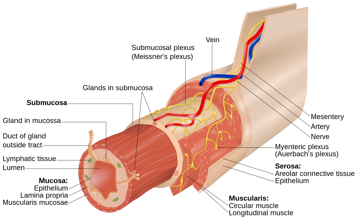
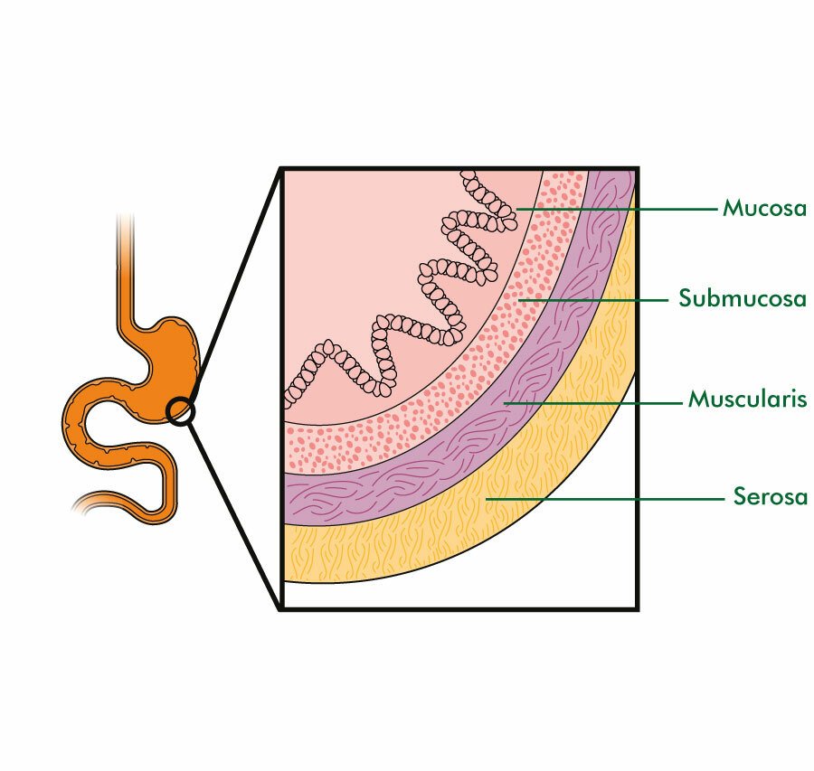
Layers of the Gastrointestinal Tract
The Gl tract from the esophagus to the anal canal is composed of four layers, or tunics. Each tunic contains a dominant tissue type that performs specific functions in the digestive process. The four tunics of the GI tract, from the inside out, are…
- The mucosa,
- Submucosa,
- Muscularis, and
- Serosa
1. Mucosa.
The mucosa, or inner lining of the tract, is a mucous membrane. It is composed of a layer of epithelium in direct contact with the contents of the GI tract, a layer of areolar connective tissue called the lamina propria, and a thin layer of smooth muscle called the muscularis mucosae.
Contractions of the muscularis mucosae create folds in the mucosa that increase the surface area for digestion and absorption. The mucosa also contains prominent lymphatic nodules that protect against the entry of pathogens through the GI tract.

2. Submucosa.
The submucosa consists of areolar connective tissue that binds the mucosa to the muscularis. It contains many blood and lymphatic vessels that receive absorbed food molecules. Also located in the submucosa are networks of neurons that are subject to regulation by the autonomic nervous system (ANS) called the enteric nervous system (ENS), the “brain of the gut.” ENS neurons within the submucosa controlthe secretions of the organs of the GI tract.
3. Muscularis.
As its name implies, the muscularis of the GI tract is a thick layer of muscle. In the mouth, pharynx, and upper esophagus, it consists in part of skeletal muscle that produces voluntary swallowing. Skeletal muscle also forms the external anal sphincter, which permits voluntary control of defecation. Recall that a sphincter is a thick circle of muscle around an opening.
In the rest of the tract, the muscularis consists of smooth muscle, usually arranged as an inner sheet of circular fibers and an outer sheet of longitudinal fibers. Involuntary contractions of these smooth muscles help break down food physically, mix it with digestive secretions, and propel it along the tract. ENS neurons within the muscularis control the frequency and strength of its contractions.
4. Serosa and peritoneum.
The serosa, the outermost layer around organs of the GI tract below the diaphragm, is a membrane composed of simple squamous epithelium and areolar connective tissue. The serosa secretes a slippery, watery fluid that allows the tract to glide easily against other organs.
The serosa is also called the visceral peritoneum that the peritoneum is the largest serous membrane of the body. The parietal peritoneum lines the wall of the abdominal cavity, the visceral peritoneum covers organs in the cavity.
Read more: