Liver, pancreas and gallbladder-The course is designed for the basic understanding of anatomical structures and physiological functions of human body, musculoskeletal system, digestive system, respiratory system; cardiovascular system; urinary system, endocrine system, reproductive system, nervous system, hematologic system, sensory organs, integumentary system, and immune system.The aim of the course is to acquire knowledge and skills regarding anatomy and physiology.
Liver, pancreas and gallbladder
The liver is the largest gland in the body. The liver regulates the chemical composition of the blood in numerous ways. In addition, the liver produces and secretes bile, which is stored and concentrated in the gallbladder prior to its discharge into the duodenum. The pancreas produces pancreatic juice, an exocrine secretion containing bicarbonate and important digestive enzymes which discharge into the duodenum through the common duct.
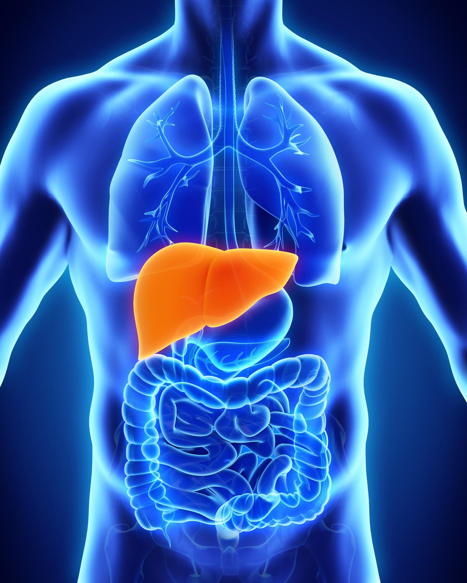
The liver is the largest gland in the body, weighing between 1 and 2.3 kg and is covered by Glisson’s capsule. It is situated in the upper part of the abdominal cavity occupying the greater part of the right hypochondriac region, part of the epigastric region and extending into the left hypochondriac region. Its upper and anterior surfaces are smooth and curved to fit the under surface of the diaphragm. Its posterior surface is irregular in outline..
(Ref:-Ross and wilson-, 9th edition, P-307.)
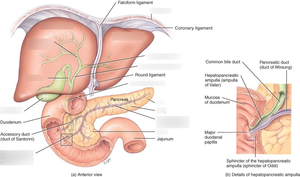
The falciform ligament is a peritoneal structure that courses between the left and right lobes of the liver and the anterior abdominal wall. Hepatic cells (hepatocytes) of the liver produce bile that is transported by a duct system to the gallbladder for storage. Hepatocytes form hepatic plates that are one to two cells thick. The plates are separated from each other by large capillary spaces called sinusoids.
The four lobes of the liver are as follows.
- Right lobe. Positioned to the right of the inferior vena cava and gallbladder.
- Left lobe. Positioned to the left ligamentum teres.
- Quadrate lobe. Positioned posterior to the portal triad. handsome
- Caudate lobe. Positioned anterior to the portal triad.
Functionally, the quadrate and caudate lobes are part of the left lobe because they are supplied by the left hepatic artery, drained by the left branch of the portal vein, and deliver bile via the left bile duct.

Microscopic structures of liver
Microscopically, the liver consists of several components:
- Hepatocytes (hepat = liver, cytes =cells),
These are the major functional cells of the liver that perform metabolic, secretory, and endocrine functions.
- Bile canaliculi
These are small ducts between hepatocytes that collect bile produced by the hepatocytes. From bile canaliculi, bile passes into bile ducts. The bile ducts merge and eventually form the larger right and left hepatic ducts, which unite and exit the liver as the common hepatic duct.
The common hepatic duct joins the cystic duct (cystic =bladder) from the gallbladder to form the common bile duct. From here, bile enters the small intestine to participate in digestion. When the small intestine is empty, the sphincter around the common duct at the entrance to the duodenum closes, and bile backs up into the cystic duct to the gallbladder for storage.
- Hepatic sinusoids.
These are highly permeable blood capillaries between rows of hepatocytes that receive oxygenated blood from branches of the hepatic artery and nutrient-rich deoxygenated blood from branches of the hepatic portal vein. Recall that the hepatic portal vein brings venous blood from the gastrointestinal organs into the liver. Hepatic sinusoids converge
and deliver blood into a central vein. From central veins the blood flows into the hepatic veins, which drain into the inferior vena cava. Also present in the hepatic sinusoids are fixed phagocytes called stellate reticuloendothelial (Kupffer) cells, which destroy worn-out white and red blood cells. bacteria, and other foreign matter in the venous blood draining from the gastrointestinal tract.
(Ref.-J. TORTORA, The essentials of anatomy and physiology, 8th edition, P-499)
Organs associated with the liver
| Superiorly and anteriorly | Diaphragm and anterior abdominal wall. |
| Inferiorly | Stomach, bile ducts, duodenum, hepatic flexure of the colon, right kidney and adrenal gland |
| Posteriorly | Oesophagus, inferior vena cava, aorta, gallbladder, vertebral column and diaphragm. |
| Laterally | Lower ribs and diaphragm |
Functions of liver
The liver performs many other vital functions in addition to the secretion of bile and bile salts and the phagocytosis of bacteria and dead or foreign material by the Kupffer cells. Many of these are related to metabolism and are discussed in however, the other vital functions of the liver include the following:
- Carbohydrate metabolism. The liver is especially important in maintaining a normal blood glucose level. When blood glucose is low, the liver can break down glycogen to glucose and release glucose into the bloodstream. When blood glucose is high, as occurs just after eating a meal, the liver converts glucose to glycogen and triglycerides for storage.
- Lipid metabolism. Hepatocytes store some triglycerides; break down fatty acids to generate ATP for providing energy
- Protein metabolism. Hepatocytes remove the amino group (-NH2) from amino acids so that the amino acids can be used for ATP production or converted to carbohydrates or fats. Hepatocytes also synthesize most plasma proteins, such as globulins, albumin, prothrombin, and fibrinogen.
- Processing of drugs and hormones. The liver can detoxify substances such as alcohol or secrete drugs such as penicillin, erythromycin, and sulfonamides into bile. It can also inactivate thyroid hormones and steroid hormones such as estrogens and aldosterone.
- Excretion of bilirubin. Bilirubin, derived from the haeme of aged red blood cells, is absorbed by the liver from the blood and secreted into bile. Most of the bilirubin in bile is metabolized in the small intestine by bacteria and eliminated in feces
- Storage of vitamins and minerals. In addition to storing glycogen, the liver stores certain vitamins (A, D, E, and K) and minerals (iron and copper), which are released from the liver when needed elsewhere in the body
- Activation of vitamin D. The skin, liver, and kidneys participate in synthesizing the active form of vitamin D
Shortly
(Ref. J. TORTORA. The essentials of anatomy and physiology. 8th edition, P-500.501)
Functions of liver
The liver is the largest chemical factory in the body and performs a variety of vital functions. The vital functions of the liver include the following
1. Production and secretion of bile
2. Metabolism of carbohydrate, fat and protein
3. Storage and distribution of glycogen, vitamin A, D, B, and also iron.
4. Production of plasma protein
5. Detoxification; liver is able to destroy or modify waste products and toxic substances in the body
6. Manufacturing of prothrombin & fibrinogen essential for the clotting of blood. etc
(Ref: P. Evelyn, Anatomy & Physiology for nurses, 16 edition, P-245, 246)
Read more: