Heart, chambers and the valves of the heart-The course is designed for the basic understanding of anatomical structures and physiological functions of human body, musculoskeletal system, digestive system, respiratory system; cardiovascular system; urinary system, endocrine system, reproductive system, nervous system, hematologic system, sensory organs, integumentary system, and immune system.The aim of the course is to acquire knowledge and skills regarding anatomy and physiology.
Heart, chambers and the valves of the heart
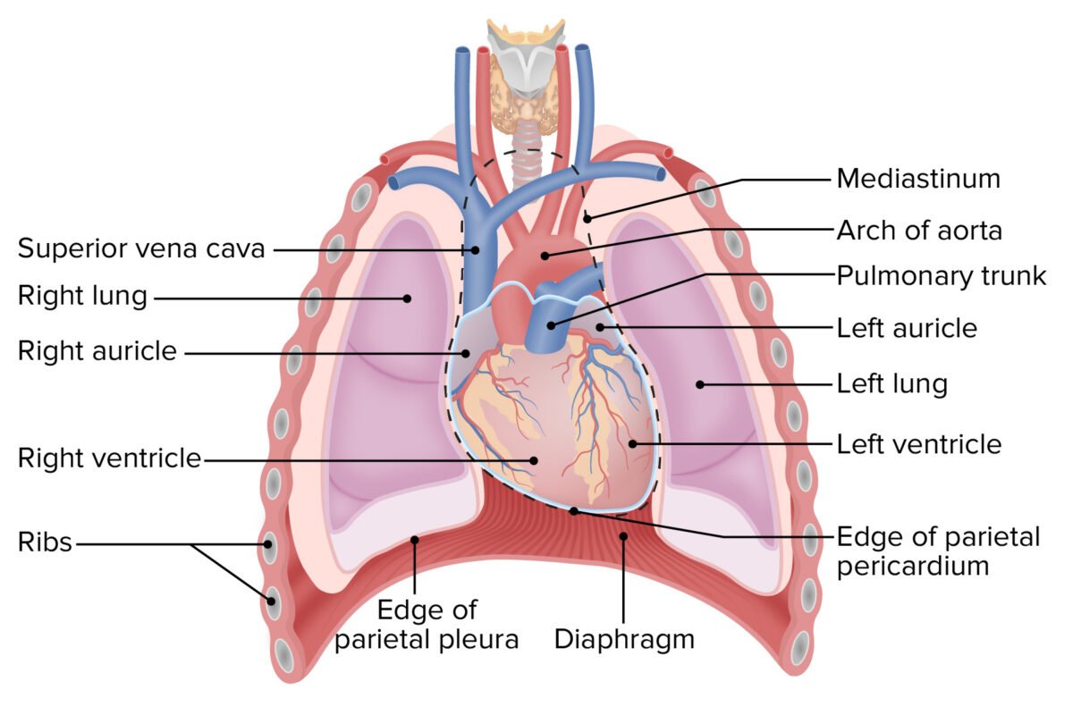
Heart: The heart is a hollow muscular organ present only in all vertebrates responsible for pumping blood through a closed blood vessels by repeated & rhythmic contractions. The heart is situated between the two lungs in the thoracic cavity, with about two-thirds of its mass lying to the left of the body’s midline (Figure 5.13). Your heart is about the size of your closed fist.
Heart’s position in thorax
In mediastinum – behind sternum and pointing left, lying on the diaphragm It weighs 250-350 gm (about 1 pound)
Chambers of the heart:
The human heart possesses four muscular chambers two chambers on the left side, left armium and let ventricle, and two chambers on the right side; right atrium and right ventride. The left and righ chambers are separated by muscle known as septum so that coxygenated blood does not comittine wit deoxygenated blood.
Four chambers of heart are –
- Left atrium.
- Left ventricle.
- Right atrium
- Right ventricle
Actually, the heart has two separate pumps –
- A right heart that pumps blood through the lungs and contains deoxygenated blood.
- A left heart that pumps blood through the peripheral organs contains oxygenated blood.
Characteristics of the atrium and ventricle-
- The ventricle has a larger volume of space than theatrium.
- The ventricle has a thicker wall than the atrium.
- The left ventricle is thicker and more muscular than the right ventricle because this chamber pumps blood under higher pressure to the entire body
Layers of heart muscle
There are three layers of heart muscle as
1. Epicardium = visceral layer of serous pericardium( outer layer)
2. Myocardium = the muscle( middle layer)
3. Endocardium lining the chambers( inner layer)
Nerve Supply to the heart
Heart is supplied by nerves arising from superficial and deep cardiac plexus. Nerves arising from these plexuses run along the coronary arteries and supply the heart. These contain sympathetic and parasympathetic nerves
Sympathetic nerves are derived from upper 4 or 5 thoracic segments (T1-T4) of spinal cord Parasympathetic nerves are derived from vagus (X) which arise from the dorsal nucleus of vagus (medulla oblongata)
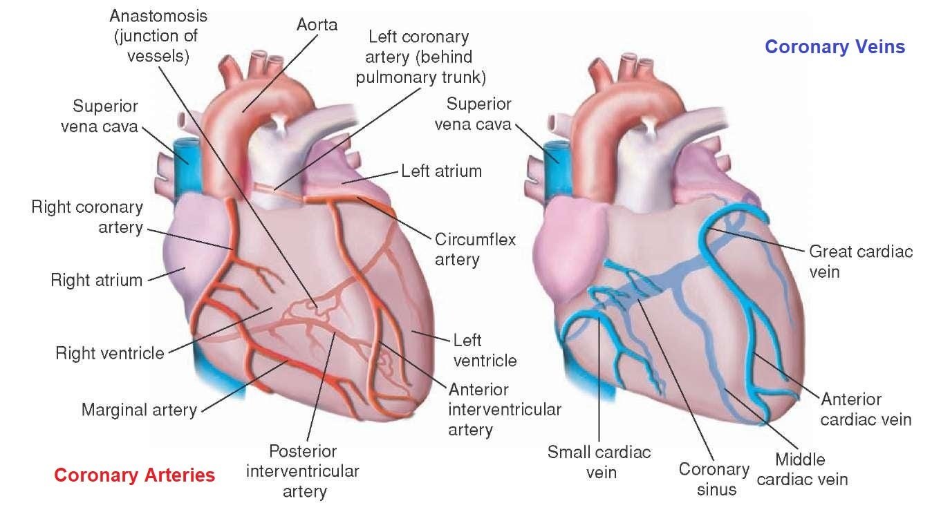
Blood supply to the heart
The heart receives its own supply of blood from the coronary arteries Two major coronary arteries branch off from the aorta near the point where the aorta and the left ventricle meet These
arteries and their branches supply all parts of the heart muscle with blood
The function of the heart
1. The heart functions as strong muscular pump to:
a. Collect deoxygenated blood (lacking oxygen) from the rest of the body Then, this blood is pumped from the heart to the lungs to enriched with oxygen
b. Collect oxygenated blood (enriched with oxygen) from the lungs. Then this blood is pumped out of the heart to be transported throughout the body
2. The heart also plays a role in the human circulatory system to:
a. Transport nutrients and oxygen to the body
b. Transport excretory products such as carbon dioxide, urea and water from the body cells to be removed from the body
(Ref: Guyton and Hall, 12 ed. P-101.)
Valves of the heart:
| Valves | location |
| Mitral or Bi-cuspid valve | Between left atrium & left ventricle |
| Tricuspid valve | Between right atrium & right ventricle |
| Aortic valve | Between left ventricle & aorta |
| Pulmonary valve | Between right ventricle & pulmonary artery |
The aorta is the largest artery in the human body, originating from the left ventricle of the heart and extending down to the abdomen, where it bifurcates into two smaller arteries (the common iliac arteries).
The aorta distributes oxygenated blood to all parts of the body through the systemic circulation and aorta is like a tube about a foot long and just over an inch in diameter. The aorta is divided into four sections.
- The ascending aorta rises up from the heart and is about 2 inches long. The coronary arteries branch off the ascending aorta to supply the heart with blood.
- The aortic arch curves over the heart, giving rise to branches that bring blood to the head, neck, and arms.
- The descending thoracicarts travels down through the chest. Its small branches supply blood to the ribs and some chest structures.
- The abdominal aurt begins at the diaphragm, splitting to become the paired iliac arteries in the lower abdomen. Most of the major organs receive blood from branches of the abdominal aorta.
The arch of aorta
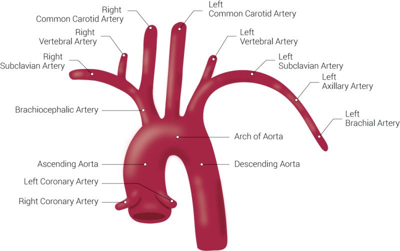
The arch of the Aorta lies within the mediastinum. The arch of aorta branches off of the first portion of the aorta, or ascending aorta. Three major arteries onginate from the aortic arch the brachiocephalic artery, which supplies blood to the brain and head; the left common carotid artery and the left subclavian artery