Today our topic of discussion is Ultrasonography of Medical Surgical Procedures.
Ultrasonography of Medical Surgical Procedures
ULTRASONOGRAPHY
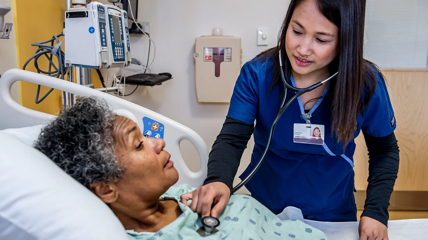
Ultrasonic waves (sound waves too high in frequency for a human ear to detect) are used diagnostically to asses various body structures. The waves are directed at the org or structure, and as they vibrate back from the target, they are transducer into oscilloscope tracing, Sonography may be used in conjunction with other pulmonary diagnostic procedures such as thoracentesis or pleural biopsy to asse fluid or fibrotic abnormalities.
Purpose
- Ultrasonography is especially helpful and very accurate in detecting the amount and location of 50 ml or less of pleural fluid.
- In comparison, a positive detection b chest radiography requires at least 500 ml. of liquid If the technique is used in combination with thoracentesis, the ultrasonographer can determine the best location for the needle placement as well as the depth of the fluid
- Ultrasonography facilitates obtaining an adequate amount of fluid for laboratory analysis without unnecessary punching and probing (Fig. 29.31).
Preoperative Care
- No special care is required before ultrasonography
- Explain the purpose of the test and what to expect
- A gel is applied to the skin, a transducer (a device that changes reflected high-frequency sound to electrical energy) is moved on the skin surface above the target organ Inform the client that the procedure is painless and fairly quick
- Procedure: Alung angiogram is administered by inserting thin tube, or catheter into a vein leading to the lungs. This tube is then guided to the area that requires studying after which the iodine is injected in order to provide a contrasting color of the veins on the final X-ray.
- During the procedure, you are probably going to be asked to put on a lead gown to protect the genital and pelvic areas from X-ray exposure and a round cylinder or rectangular box that captures the images will be passed over the targeted area (Fig. 29.32).
- The clients are placed on an X-ray table in the supine position
- Electrocardiography electrodes are attached for cardiac monitoring.
- The catheter is placed into the femoral vein and passed into the inferior vena cave With fluoroscopic visualization, the catheter is advanced to the right atrium and the right ventricle
- The catheter is manipulated into the main pulmonary artery, where the dye is injected
- X-ray films of the chest are immediately taken in sequence.
- This allows all vessels visualized by injection to be photographed.
- If filling defects are seen the contrast-filled vessels, pulmonary emboli are present
- If bronchial artery is performed, the femoral artery is cannulated instead of the vein
- During injection of dye, inform the client that he or she will feel a burning sensation and flush throughout the body.

Post-procedural Care
- Observe the catheter insertion site for inflammation, hemorrhage and hematoma Assess the client’s vital signs for evidence of bleeding
- (decreased blood pressure, increased pulse)
- Apply cold compress to puncture site if needed to reduce swelling or discomfort Inform the client that coughing may occur after this study .
- Educate the client regarding the need for bed rest for 12 to 24 hours after the test.