
Today our topic of discussion is Pleural Biopsy. The human respiratory system is a marvel of evolution, with various components working in tandem to facilitate the vital process of respiration. The pleura, the thin membrane that envelopes the lungs, plays a pivotal role in ensuring efficient lung function. However, like all structures in the body, the pleura is susceptible to various conditions and diseases. A pleural biopsy is an invaluable procedure to diagnose and treat these ailments.
Pleural Biopsy
What is the Pleura?
The pleura is a thin, double-layered membrane that envelopes both lungs. The two layers are the:
- Visceral pleura: This covers the lungs themselves.
- Parietal pleura: This lines the inside of the chest wall.
The space between these two layers, known as the pleural space, contains a small amount of fluid known as pleural fluid. This lubricates the two surfaces, allowing them to slide against each other without friction during breathing.
Why is a Pleural Biopsy Done?
There are several reasons a healthcare provider might recommend a pleural biopsy:
- Diagnostic purposes: It can diagnose diseases such as tuberculosis, pleural tumors, or other infections.
- Pleural effusion evaluation: A buildup of excess fluid in the pleural space can be due to a plethora of reasons, including infection, cancer, heart failure, or systemic diseases like lupus. Analyzing the pleural tissue can provide insights into the underlying cause.
- Therapeutic purposes: Removing fluid from the pleural space can help alleviate symptoms like shortness of breath.
Types of Pleural Biopsies
There are several methods to conduct a pleural biopsy:
- Closed or blind pleural biopsy: Traditionally done using an Abrams or Cope needle, this method doesn’t employ imaging guidance.
- Image-guided biopsy: Ultrasound or CT-guided biopsies are done to ensure precision, especially when the target area is small or adjacent to vital structures.
- Thoracoscopic biopsy: Also known as video-assisted thoracoscopic surgery (VATS), this procedure utilizes a camera and specialized instruments inserted through small incisions in the chest wall.
Risks and Complications
As with any invasive procedure, a pleural biopsy does come with risks:
- Infection: Rarely, the biopsy site or the pleural space may become infected.
- Bleeding: There might be slight bleeding at the biopsy site or within the pleural space.
- Pneumothorax: Occasionally, air can enter the pleural space causing lung collapse. It might require a chest tube to remove the air and re-expand the lung.
- Pain: Some discomfort is normal after the procedure.
PLEURAL BIOPSY
Biopsy specimens may be taken from various respiratory rissues for examination. As mentioned previously, specimens from tracheobronchial structures may be obtained during bronchoscopy. Biopsy specimens of
scalene and mediastinal nodes may be obtained (with local anesthesia for pathologic study, culture or cytological assessment (Fig. 29.42).
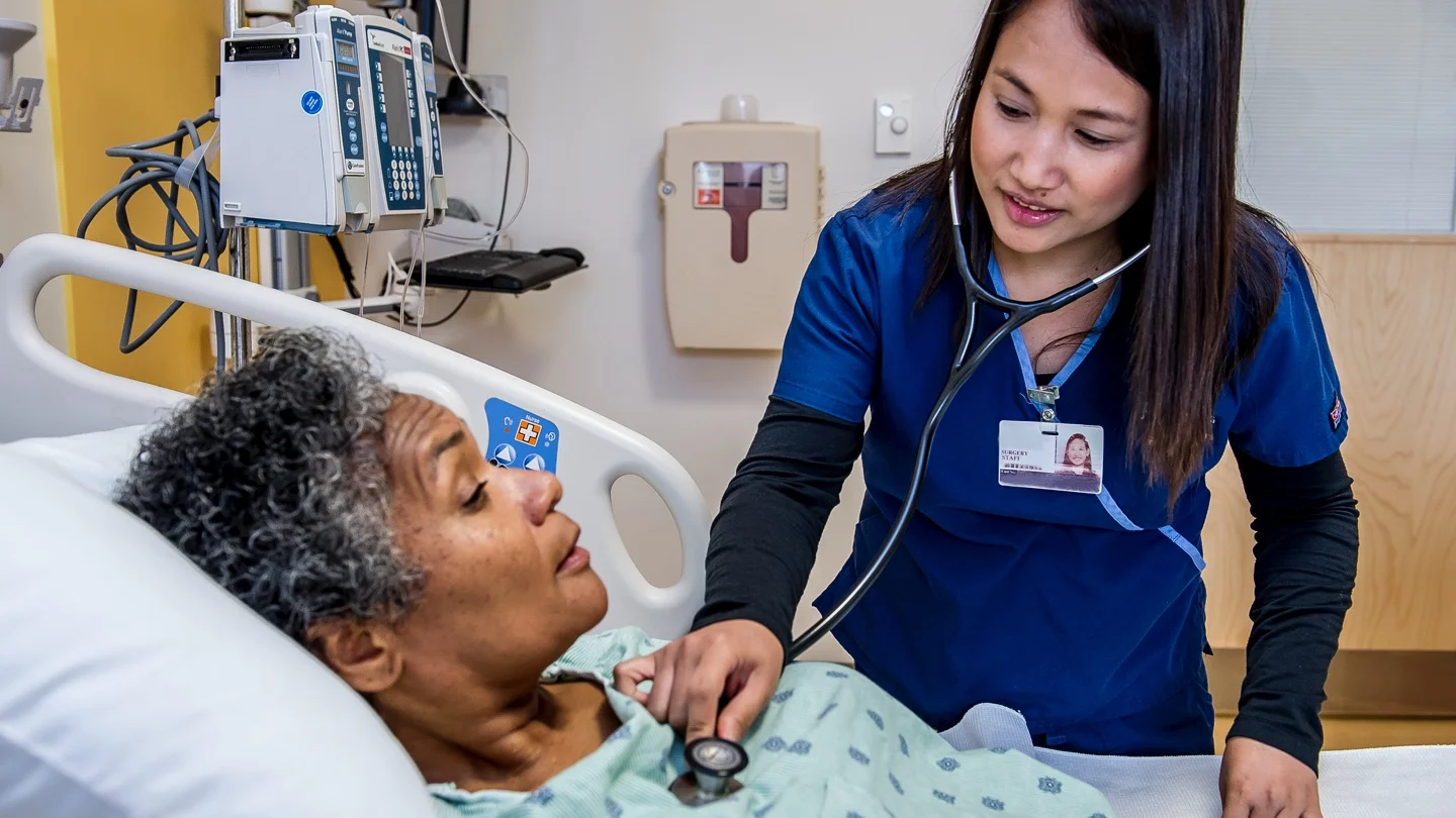
Pre-procedure Care
- Obtain inform consent, and instruct the client about the need for and purpose of the test
- Preparation and positioning of a client for pleural biopsy are similar to those for thoracentesis
- Inform the client, the test is painful, and the client must hold still
- Assist and reassure the client.
- The test takes 15-30 minutes to complete.
Procedure
- Pleural biopsies can be performed surgically through a small thoracotomy incision or during thoracentesis, with the use of a cope needle
- Needle biopsy is a relatively safe, simple diagnostic procedure that can help to determine the cause of pleural effusion
- The needle removes a small fragment of parietal pleura, which is used for microscopic cellular examination and culture
- If bacteriologic studies are needed, the biopsy specimen should be obtained before chemotherapy is begun.
Post-procedure Care
- After the biopsy procedure, observe for indications of complications (dyspnea, pallor, diaphoresis, excessive pain)
- Follow-up chest X-ray studies are usually done after the procedure.

Complications
Rare complications include temporary pain associated with intercostals nerve injury, and pneumothorax.
Results and Interpretation
Biopsy samples are sent to a pathology lab for microscopic examination:
- Benign findings: Conditions like granulomatous inflammation can hint towards infections like tuberculosis.
- Malignant findings: Presence of cancerous cells might indicate primary lung cancer or metastatic disease.
- Nonspecific findings: Sometimes, the biopsy might not provide a definitive diagnosis, necessitating further tests or repeat biopsy.
Conclusion
A pleural biopsy is a crucial tool in the diagnostic arsenal of pulmonologists and thoracic surgeons. Like any procedure, it comes with risks, but its utility in diagnosing potentially life-threatening conditions can’t be overstated.
When recommended, patients should discuss with their healthcare provider the reasons, potential risks, and benefits. As technology and medical techniques advance, pleural biopsies will likely continue to become more accurate and less invasive, but their fundamental role in diagnosis and patient care will remain vital.