
Today our topic of discussion is Intravenous Pyelography.
Intravenous Pyelography
INTRAVENOUS PYELOGRAPHY
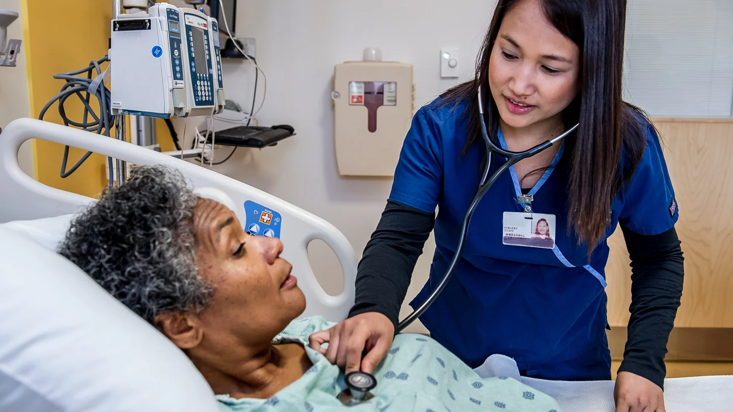
Intravenous pyelography (IVP) is also called excretory urogram. Intravenous pyelography is the roentgenographic visualization of kidneys, ureters and bladder by injecting a dye into the vascular system. It involves intravenous injection of a radiopaque dye that is filtered by the kidney and excreted through the urinary tract (Fig. 29.113).
Purpose
- To identify absence or presence, location size and configuration of kidneys, ureters and bladder
- To determine filling of the renal calices
- To detect anatomic peculiarities of the urinary system, such as horseshoe kidney, polycystic kidney. hydronephrosis, double ureters, etc.
- To find out enlargement of prostate gland, tumors, renal calculi, abnormalities of the urinary bladder, etc.
- Post-volding films, showing abnormal retention of dye,
- can indicate bladder neck obstruction.
Contraindications
A known sensitivity to iodinated contrast media is an absolute contraindication to IVP.
Preparation of the Patient
- Explain the procedure to the patient to relieve his fear and anxiety
- Obtain written consent from the patient
- Test skin to find out sensitivity to iodine compounds because radiopaque medium containing iodine, when injected intravenously, can cause anaphylactic reactions in hypersensitive patients
- Prepare the patient as for taking plain X-ray of abdomen
- Keep the patient nil by mouth after evening meal until the examination is over.
- The depletion of fluid intake allows the radiopaque dye to be more concentrated.
- When it enters the kidney, the roentgenogram is clearer.
- If the patient is receiving intravenous fluids, the rate of infusion may be slowed down, for several hours, prior to the study
- Because the kidneys are located retroperitoneally, the bowel must be cleared of gas and fecal matter, because the gas and fecal matter may cause shadows in the X-ray film
- Cathartics are usually given evening before the examination.
- If the patient is suffering from colitis or peptic ulcer, he should not be given cathartics
- The nurse must take precautions to assure the comfortm and safety of the patient
- A history of kidney damages, allergy, liver diseases, and cardiac arrhythmia are contra-indications to the use of radiopaque substances
- Keep emergency drugs, oxygen, and resuscitation equipment ready.
Commonly Used Substances for IVP
The dyes currently used are di- and tri-iodinated derivatives of benzene and pyridine.Two commonly used substances are: a. Diatrizoate sodium (Hypaque) b. Meglumine diatrizoate (Renogram)
Position of the Patient
The patient is placed in a supine position on the X-ray table.
Procedure
- The patient is placed in a supine position on the X-ray table
- Initially a KUB film is taken
- This helps assure that the bowel is clear enough to continue with the rest of the procedure.
- It also screens for calculi in the kidney, ureters or bladder The radiopaque dye is injected intravenously.
- Films are usually taken 2,5, 10,15,20,30 and 60 minutes after the dye is injected. Post-voiding films also may be taken.
- The drug should not be injected by the nurse.
- The compounds normally cause flushing of face, a feeling of warmth and a salty taste in mouth.
- These effects are transitory and do not mean that the study should be stopped.
After Care
- Sometimes, with delayed renal function, additional X-rays may be needed one or two hours later.
- If the patient is to remain on the X-ray table, during this time care must be taken to insure comfort, as much as possible
- The patient should be observed for any untoward reactions during and after the procedure
- The patient must be watched for the following complications: Anaphylactic shock, acute renal failure, electrolyte imbalance and cardiac arrhythmias
- If the patient develops any signs of allergic reaction such
- as itching, wheezing or other respiratory distress, stop the procedure immediately
- When the patient returns to the ward, observe him for reaction of the dye
- Give plenty of oral fluids to rehydrate the patient.

Recording and Reporting
- Record the vital signs before and after the procedure on the nurse’s chart
- Record any reaction observed and report to the sister/ physician.
Complications
- Anaphylactic shock
- Acute rena