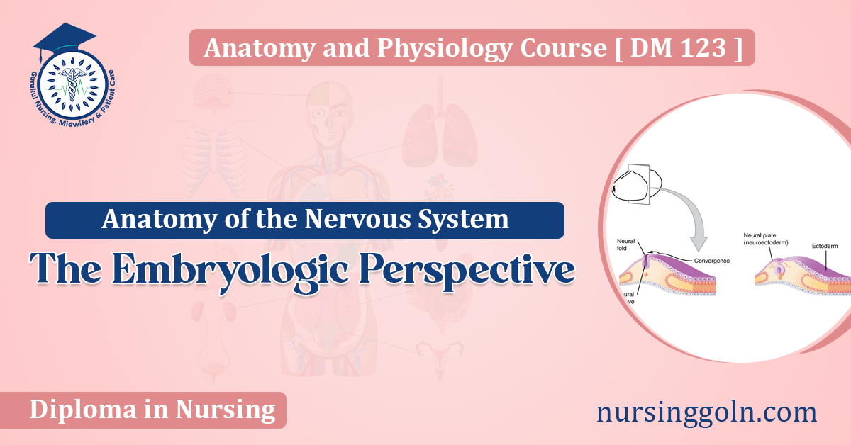
Today our topic of discussion is ” The Embryologic Perspective “. The marvel that is the human nervous system doesn’t emerge fully formed but undergoes a complex journey from embryonic development to a fully functional network of nerves and neurons. This article examines the anatomy of the nervous system through the lens of embryology, illuminating the intricate processes that guide its formation.
The Embryologic Perspective: Anatomy of the Nervous System
1. Embryogenesis and the Emergence of the Neural Plate
Embryogenesis, the process of forming a new organism, begins with the fertilized egg, or zygote. As this cell divides and specializes, by the third week of development, a structure known as the neural plate forms. This flat layer of cells is the foundation of the nervous-system.
2. Neural Tube Formation: The Birth of the CNS
Around the 22nd day of embryonic development, the edges of the neural plate start to elevate, forming the neural folds. These folds gradually move towards one another, eventually fusing to create the neural tube. This structure is a precursor to the central nervous-system (CNS), which encompasses the brain and spinal cord.
3. Neural Crest and the Advent of the PNS
As the neural tube forms, some cells, known as neural crest cells, pinch off from the top of the neural folds. These cells migrate throughout the embryo, laying the groundwork for the peripheral nervous system (PNS) and various other structures.
4. Differentiation of the Neural Tube
The rostral (head) end of the neural tube differentiates into the brain, while the caudal (tail) end forms the spinal cord. The developing brain further divides into three primary vesicles:
- Prosencephalon (Forebrain): Later differentiates into the telencephalon (giving rise to the cerebral hemispheres) and the diencephalon (evolving into the thalamus and hypothalamus).
- Mesencephalon (Midbrain): Remains relatively undivided.
- Rhombencephalon (Hindbrain): Segregates into the metencephalon (precursor to the cerebellum and pons) and the myelencephalon (leading to the formation of the medulla oblongata).
5. Ventricular System and CSF
Inside the neural tube is a small cavity that expands to form the ventricular system of the brain and the central canal of the spinal cord. These spaces are filled with cerebrospinal fluid (CSF), produced by specialized structures called choroid plexuses.
6. The Gray and White Matter
As the CNS matures, its internal structure starts to differentiate. Neuronal cell bodies group together to form the gray matter, while myelinated axonal fibers form the white matter. This distinction becomes particularly evident in the spinal cord.
7. Maturation of the Peripheral Nervous System (PNS)
Recall the neural crest cells. As they migrate and differentiate, they give rise to sensory and autonomic ganglia, adrenal medulla, and various other PNS components. The motor portion of the PNS arises from the neural tube itself.
8. The Blood-Brain Barrier
The CNS requires protection from potentially harmful substances in the bloodstream. During development, specialized capillaries form the blood-brain barrier, which strictly regulates the entry of substances into the CNS.
9. Genetic and Environmental Influences
The entire embryological journey of the nervous system is orchestrated by a combination of genetic programming and environmental factors. Any disruption, be it due to genetic mutations or external teratogens, can lead to neurodevelopmental disorders.
10. Conclusion: The Embryological Symphony
The anatomy of the nervous system, when viewed through an embryologic lens, is nothing short of a symphony. Each step, from the formation of the neural plate to the maturation of the CNS and PNS, plays a crucial note in the harmonious creation of an intricate system that governs thought, perception, emotion, and movement. Understanding this developmental journey not only offers insights into the anatomy but also provides valuable knowledge for addressing congenital and developmental disorders of the nervous system.