Today our topic of discussion is ” Microscopic Anatomy of the Kidney “. The microscopic anatomy of the kidney reveals a complex and highly organized structure, essential for its role in filtration, secretion, and reabsorption within the urinary system. At this microscopic level, the kidney is composed of an array of specialized cells and tissues that together form the functional units known as nephrons.
Each kidney contains approximately one million nephrons, and it is within these microscopic structures that blood is processed, and urine is formed. This article will explore the microscopic components of the kidney, including the structure and function of nephrons, the vascular supply, and the interstitial tissue, to provide a detailed understanding of renal physiology and pathology.
Microscopic Anatomy of the Kidney : The Urinary System
Nephrons: The Functional Units
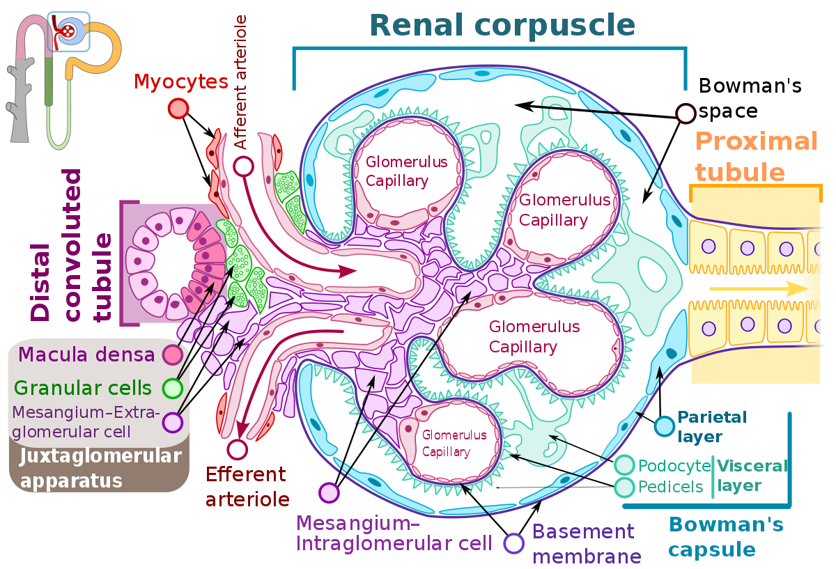
Glomerulus
- Description of the glomerular capillary tuft
- The glomerular basement membrane and its significance
- Podocytes and their foot processes
- Mesangial cells function
Bowman’s Capsule
- The inner visceral and outer parietal layer structure
- Bowman’s space and its significance infiltration
Proximal Convoluted Tubule (PCT)
- Brush border microvilli
- Mitochondria-rich cells for active transport
- Role in reabsorption and secretion
Loop of Henle
- Descending and ascending limbs
- Thin and thick segments
- Role in the countercurrent mechanism
Distal Convoluted Tubule (DCT)
- Differences in cell structure from PCT
- Role in ion exchange and acid-base balance
Collecting Duct
- Principal cells and intercalated cells
- Role in water reabsorption and urine concentration

Vascular Supply
Afferent and Efferent Arterioles
- Regulation of glomerular blood flow
- Juxtaglomerular apparatus and its role in blood pressure regulation
Peritubular Capillaries and Vasa Recta
- Nutrient supply and gas exchange
- Role in the countercurrent exchange system
Interstitium
Interstitial Cells
- Functions of interstitial fibroblasts and immune cells
- Extracellular matrix composition
Interstitial Fluid
- Compositional differences from plasma
- Role in the transport of solutes
Juxtaglomerular Apparatus
Macula Densa
- Sensing sodium concentration in the DCT
- Signaling mechanisms to juxtaglomerular cells
Juxtaglomerular Cells
- Renin secretion in response to blood pressure changes
Extraglomerular Mesangial Cells
- Structural support and signaling within the JGA

Renal Corpuscle and Filtration Barrier
Filtration Slits and Membranes
- Role in preventing proteinuria
- Charge and size selectivity
Podocyte Injury and Glomerular Diseases
- Correlation with diseases like diabetes and hypertension
Tubular Transport Mechanisms
Active and Passive Transport
- Energy-dependent and energy-independent mechanisms
- Transporters and channels involved
Tubuloglomerular Feedback
- Fine-tuning of glomerular filtration rate
![]()
Pathophysiology of Microstructures
Acute Kidney Injury
- Cellular responses to injury and potential for recovery
Chronic Kidney Disease
- Microstructural changes over time
- Impact on renal function
Advances in Microscopic Imaging
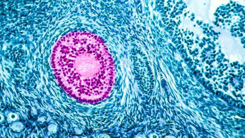
Light Microscopy and Staining Techniques
- Histological examination of kidney tissues
Electron Microscopy
- Detailed visualization of ultrastructural changes
Confocal Microscopy
- Live imaging of kidney cells and processes
Regenerative Medicine and the Kidney
Stem Cells and Kidney Repair
- Potential for regenerative therapies
Tissue Engineering
- Artificial kidney and tissue constructs
Conclusion
The microscopic anatomy of the kidney offers a fascinating window into the complex functions of this vital organ. Understanding these microstructures is critical for comprehending how the kidney operates under normal physiological conditions and how it responds to pathology. As research and technology progress, the detailed study of kidney microanatomy continues to shed light on new diagnostic markers and therapeutic targets for renal disease, with the potential to improve outcomes for patients around the world.