All About Shigella – Basic microbiology, parasitology, and immunology; nature, reproduction, growth, and transmission of common microorganisms and parasites in Bangladesh; prevention including universal precaution and immunization, control, sterilization, and disinfection; and specimen collections and examination. Students will have an understanding of common organisms and parasites caused human diseases and acquire knowledge about the prevention and control of those organisms.
All About Shigella
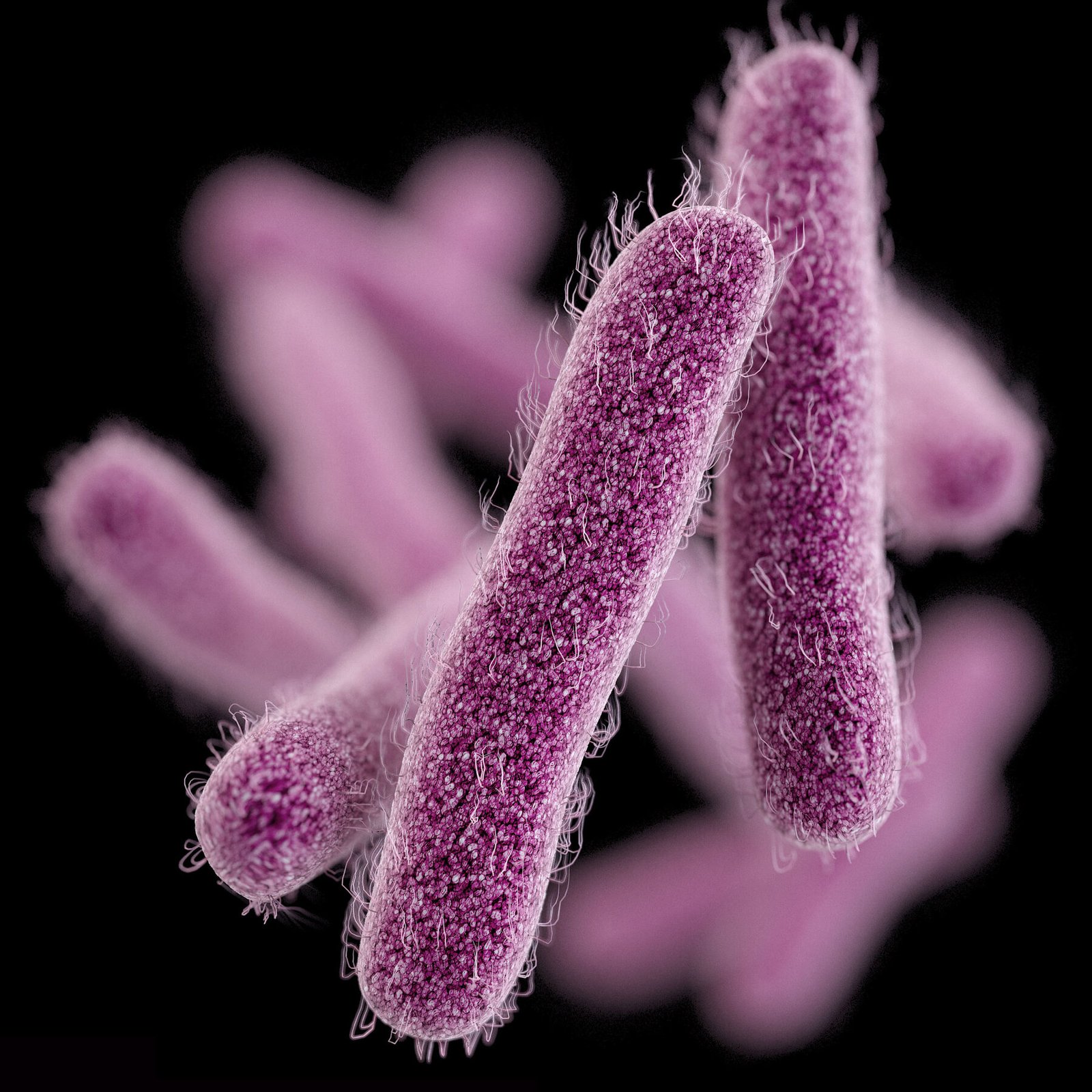
Important Properties of Shigella
- Gram-negative rods
- Non-lactose fermenter
- Form colourless colonies on Mac. Conkey’s agar media.
- Non-motile.
- Fimbriae present (In case of Sh. flexneri).
- Aerobes or facultative anaerobes
- Non-spore forming and non-capsulated
- True intestinal.

Disease Caused by Shigella Species:
➤ Shigella species cause bacillary dysentery (Shigellosis / Enterocolitis). It mostly occurs in children and it is the leading cause of infant death in developing countries like Bangladesh.
➤ Bacillary dysentery is characterized by frequent painful passage of stools containing blood and mucus.
Classification of Shigella:
All shigella have O antigens (polysaccharide) in their cell walls, and these antigens are used to divide the genus into four groups: A, B, C, and D.
| Groups | Species |
| Group A. Mannitol non-fermenters | Shigella dysenteriae |
| Group B. Mannitol fermenters. Serologically not distinct | Shigella flexneri. |
| Group C. Mannitol fermenters. Serologically distinct | Shigella boydii |
| Group D. Late lactose fermenters. Mannitol fermenters. Serologically distinct. | Shigella sonnei. |
Pathogenesis of Shigellosis / Bacillary Dysentery:
➤ Route of entry: Fecal-oral route (via fingers, flies, food, faeces)
➤ Infective dose: 10 to 100 organisms are sufficient to produce disease.
Steps of Pathogenesis
Organisms reach the terminal ileum and colon
↓
Attachment of organisms to the surface of epithelial cells
↓
Shigellae penetrate the cells and multiply there.
↓
Then they spread laterally into adjacent cells as well as to the lamina propria where multiplication and bacterial colonization take place.
↓
Liberate toxin (and exotoxin called ‘shiga toxin’).
↓
Necrosis of surface epithelial cells. Lamina propria and submucosa develop an acute inflammatory reaction with formation of micro-abscesses on mucosal surface predominantly polymorphonuclear leucocytes, along with capillary thrombosis.
↓
The necrotic epithelia become soft and friable and are sloughed out in patches forming superficial ‘snail-tract’ or serpiginous ulcers, which leads to bleeding from mucosa
↓
Endotoxin irritates the bowel causing diarrhoea.
↓
Dysentery
Lab. Diagnosis of Bacillary Dysentery:
Principle: It is based on demonstration of causative agent by microscopic examination. Isolation and identification is done by bacteriological techniques. Biochemical and serological tests are also helpful.
➤ Specimen collection:
- Fresh stool
- Rectal swab
➤ Microscopic examination: Findings
- Plenty of pus cells
- Macrophage
- Some RBC
➤ Isolation and identification from culture: Culture is done in-
- Mac Conkey’s agar media or
- DCA media or
- Salmonella-Shigella agar media
(At 37°C temperature for 24-48 hours)
➤ Route stool examination:
- Physical: Odourless. Mucus & blood in stool
- Chemical: Alkaline stool
➤ Serological tests:
- Co-agglutination test to detect the endotoxin
- Slide agglutination test for confirmation of shigella and for determination of its group.