Anthrax/ Bacillus anthracis – Basic microbiology, parasitology, and immunology; nature, reproduction, growth, and transmission of common microorganisms and parasites in Bangladesh; prevention including universal precaution and immunization, control, sterilization, and disinfection; and specimen collections and examination. Students will have an understanding of common organisms and parasites caused human diseases and acquire knowledge about the prevention and control of those organisms.
Anthrax/ Bacillus anthracis
Properties of Bacillus Anthracis:
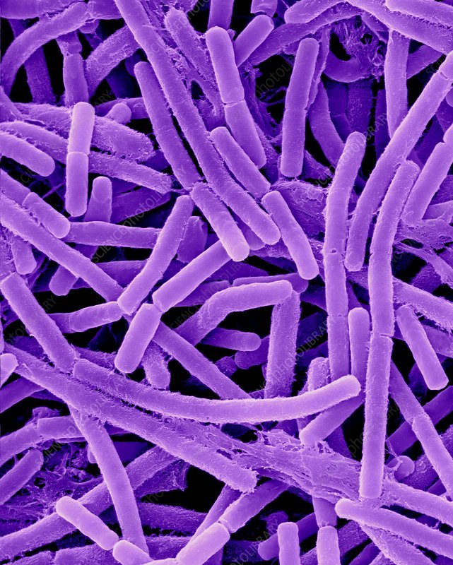
- Gram positive, spore forming rods.
- Rectangular in shape and arranged in chains. (Box car appearance)
- Possess antiphagocytic capsule, which is composed of polypeptide (polymer of D- glutamate)
- Non-motile, non-acid-fast.
- Spores are usually oval, sub-terminal or central in position and are of same diapety as the bacillary body.
Remember:
- Capsules of most bacteria consist of polysaccharide.
- In Bacillus anthracis, the capsule is a polypeptide (polymerized D-glutamate).
- In some strains of Streptococcus pyogenes, capsule is composed of hyaluronic acid.
Virulence Factors of Bacillus Anthracis:
➤ Anthrax toxin:
It is an exotoxin that has 3 components –
- Protective antigen: Protein
- Oedema factor
- Lethal factor
➤ Capsular polypeptide
Pathogenesis of Anthrax:
Bacillus anthracis invades the host tissue
↓
Production of anthrax toxin
↓
Anthrax toxin has 3 components:
- Protective antigen
- Edema factor
- Lethal factor
↓
Protective antigen forms spores in the human cell membrane
↓
Edema factor and lethal factor enter into cell
↓
Cleavage of the phosphokinase
that activates mitogen activated protein kinase (MAPK) signal transduction pathway) by lethal factor
↓
Inhibits pathway that control the growth of human cell
↓
Inhibition of cell growth
↓
Anthrax
Laboratory Diagnosis of Anthrax:
Principle:
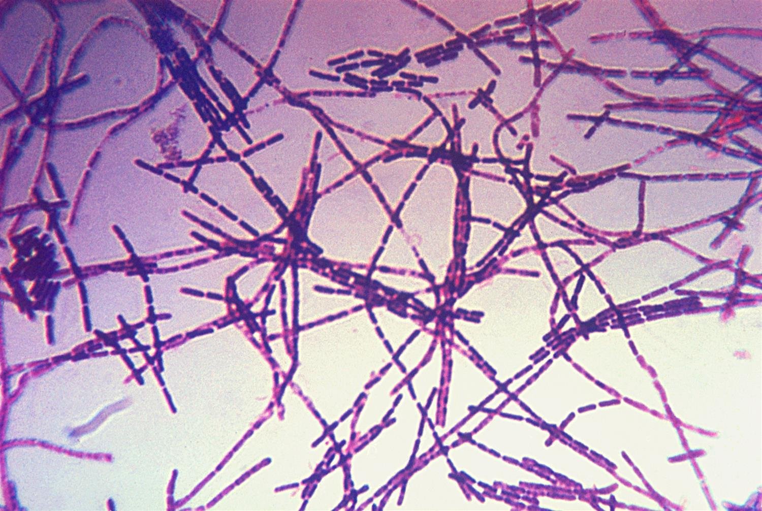
Diagnosis of anthrax is based on demonstration of the organism by microscopic examination with Mc- Fadyean-Methylene blue- staining. Serological tests are also helpful.
Steps:
- Specimen collection: Fluid, Pus. Blood, Sputum
- Microscopic examination:
✓ Stained by Me. Fadyean blue reaction
✓ Reaction is positive (large, Gram positive rods in chains).
- Isolation and identification from culture:
✓ Nutrient agar.
✓ Blood agar (Finding: aerobic colonies are formed)
- Ascoli’s thermo-precipitin test.
- Serology
✓ In vivo: Neutralizing test.
✓ In vitro: Gel diffusion test