Bone ossification – The course is designed for the basic understanding of anatomical structures and physiological functions of human body, musculoskeletal system, digestive system, respiratory system; cardiovascular system; urinary system, endocrine system, reproductive system, nervous system, hematologic system, sensory organs, integumentary system, and immune system.
The aim of the course is to acquire knowledge and skills regarding anatomy and physiology.
Bone ossification
Ossi = bone
fication = making
Ossification means bone making process.
Definition: The process by which bone forms is called ossification.
The two methods of bone formation, which both involve the replacement of a preexisting connective tissue with bone, do not lead to differences in the structure of mature bones, but are simply different methods of bone development.
- In the first type of ossification, called intramembranous ossification, bone forms directly within mesenchyme arranged in sheet-like layers that resemble membranes.
- In the second type, endochondral ossification, bone forms within hyaline cartilage that develops from mesenchyme
Bone formation occurs in four principal situations as:
(1) The initial formation of bones in an embryo and fetus.
(2) The growth of bones during infancy childhood, and adolescence until their adult sizes are reached.
(3) The remodeling of bone (replacement of old bone tissue by new bone tissue throughout life) and
(4) The repair of fractures (breaks in bones) throughout life
(Ref:- J. Tortora, The essentials of anatomy and physiology, 8th edition, P-123)
Intramembranous Ossification
Intramembranous ossificationis the simpler of the two methods of bone formation The flat bones of the skull, mandible (lower jawbone) and part of the clavicle (collar bone) are formed in this way Also, the “soft spots” that help the fetal skull pass through the birth canal later harden as they undergo intramembranous ossification which occurs as follows
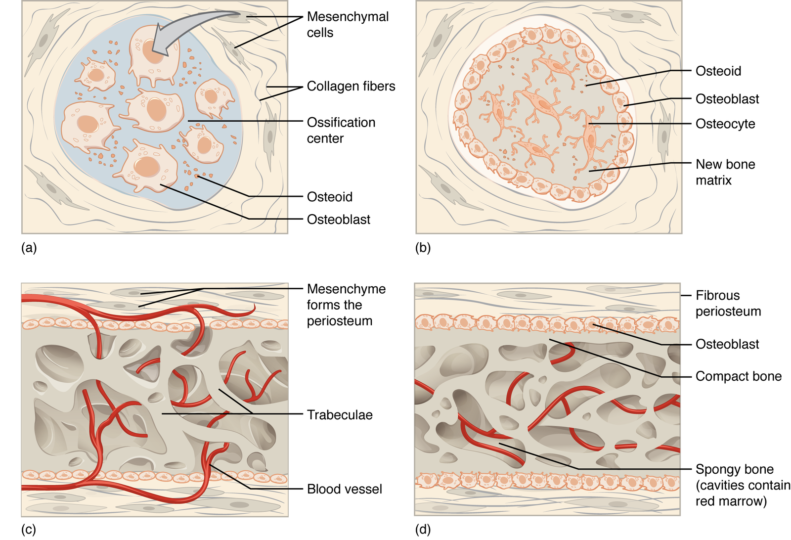
- Development of the ossification center: At the site where bone will develop, called the ossification center, mesenchymal cells cluster together and differentiate, first into osteogenic cells and then into osteoblasts. Osteoblasts secrete the organic extracellular matrix of bone
- Calcification: Next, the secretion of extracellular matrix stops and the cells, now called osteocytes, lie in lacunae and extend their narrow cytoplasmic processes into canalicult that radiate in all directions. Within a few days, calcium and other mineral salts are deposited and the extracellular matris hardens or calcifies (calcification)
- Formation of trabeculae: As the bone extracellular matrix forms, it develops into trabeculae that fuse with one another to form spongy bone Blood vessels grow into the spaces between the trabeculae Connective tissue that is associated with the blood vessels in the trabeculae differentiates into red bone marrow
- Development of the periosteum: In conjunction with the formation of trabeculae. mesenchyme condenses at the periphery and develops into the periosteum. Eventually, a thin layer of compact bone replaces the surface layers of the spongy bone, but spongy bone remains in the center.
- Development of ossification center: osteoblasts secrete organic extracellular matrix
- Calcification: calcium and other mineral salts are deposited and extracellular matrix calcifies (hardens)
- Formation of trabeculae: extracellular matrix develops into trabeculae that fuse to form spongy bone
- Development of the periosteum: mesenchyme at the periphery of the bone develops into the periosteum
Endochondral Ossification
The replacement of cartilage by bone is called endochondral ossification. Most bones of the body are formed in this way, but this type of ossification is best observed in a long bone:
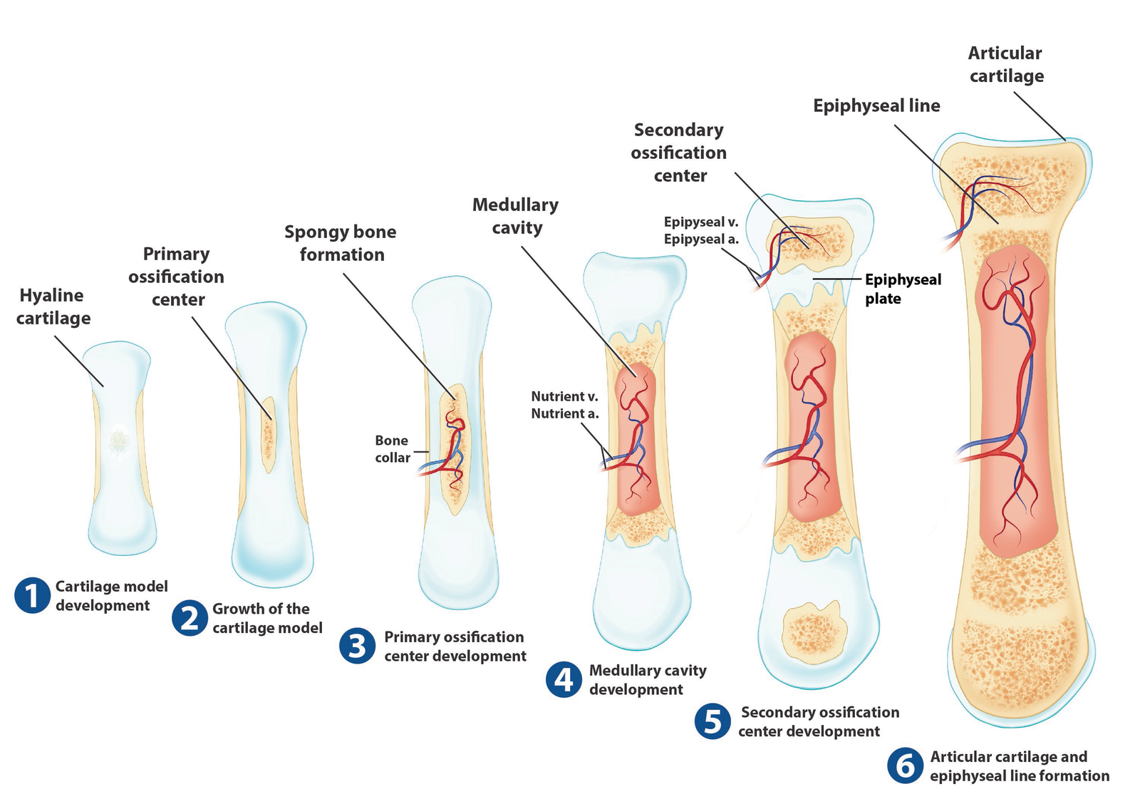
- Development of the cartilage model. At the site where the bone is going to form, mesenchymal cells crowd together in the shape of the future bone and then develop into chondroblasts. The chondroblasts secrete cartilage extracellular matrix, producing a cartilage model consisting of hyaline cartilage. A membrane called the perichondrium develops around the cartilage model.
- Growth of the cartilage model, Once chondroblasts become deeply buried in cartilage extracellular matrix, they are called chondrocytes. As the cartilage model continues to grow, chondrocytes in its mid-region increase in size and the surrounding extracellular matrix begins to calcify. Other chondrocytes within the calcifying cartilage die because nutrients can no longer diffuse quickly enough through the extracellular matrix.
- Development of the primary ossification center. Primary ossification proceeds inwand from the external surface of the bone. A nutrient artery penetrates the perichondrium and the calcifying cartilage model in the midregion of the cartilage model, stimulating osteogenic cells in the perichondrium to differentiate into osteoblasts. Once the perichondrium starts to form bone, it is known as the periosteum. Near the middle of the model, blood vessels grow into the disintegrating calcified cartilage and induce growth of a primary ossification center, a region where bone tissue will replace most of the cartilage. Osteoblasts then begin to deposit bone extracellular matrix over the remnants of calcified cartilage, forming spongy bone trabeculae. Primary ossification spreads toward both ends of the cartilage model.
- Development of the medullary (marrow) cavity, As the primary ossification center grows toward the ends of the bone, osteoclasts break down some of the newly formed spongy bone trabeculae. This activity leaves a cavity, the medullary (marrow) cavity, in the diaphysis (shaft). Most of the wall of the diaphysis is replaced by compact bone.
- Development of the secondary ossification centers. When blood vessels enter the epiphyses, secondary ossificationcenters develop, usually around the time of birth. Bone formation is similar to that in primary ossification centers except that spongy bone remains in the interior of the epiphyses (no medullary cavities are formed there). Secondary ossification proceeds ourward from the center of the epiphysis toward the outer surface of the bone.
- Formation of articular cartilage and the epiphysealplate. The hyaline cartilage that covers the epiphyses becomes the articular cartilage. Prior to adulthood, hyaline cartilage remains between the diaphysis and epiphysis as the epiphyseal (growth) plate, which is responsible for the lengthwise growth of long bones.
- Development of cartilage model: mesenchymal cells develop into chondroblasts, which form the cartilage model
- Growth of cartilage model: growth occurs by cell division of chondrocytes
- Development of primary ossification center: in this region in the diaphysis, bone tissue replaces most of the cartilage
- Development of the medullary (marrow) cavity: bone breakdown by osteoclasts forms the medullary cavity
- Development of secondary ossification centers: these occur in the epiphyses of the bone
- Formation of articular cartilage and epiphyseal plate: both structures consist of hyaline cartilage