Classification of Hydrocephalus – This course is designed to understand the care of pregnant women and newborn: antenatal, intra-natal and postnatal; breast feeding, family planning, newborn care and ethical issues, The aim of the course is to acquire knowledge and develop competencies regarding midwifery, complicated labour and newborn care including family planning.
Classification of Hydrocephalus
Hydrocephalus
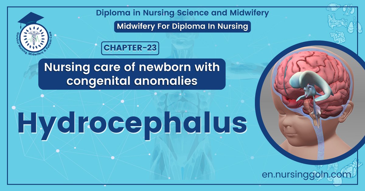
Hydrocephalus is the abnormal accumulation of cerebrospinal fluid (CSF) in the intracranial spaces. It occurs due to imbalance between production or absorption of CSF or due to obstruction of the CSF pathways.
Or
Hydrocephalus is a condition in which there is an accumulation of cerebrospinal fluid (CSF) within the brain.

Fig: Hydrocephalus
Etiology of hydrocephalus
Congenital Hydrocephalus
It occurs due to the followings conditions:
1. Intrauterine infections-mainly in rubella, toxoplasmosis, cytomegalovirus.
2. Congenital brain tumor obstructing the CSF flow.
3. Intracranial hemorrhage.
4. Congenital malformations like aqueduct stenosis, Arnold- Chiari malformation (displacement of the brainstem and cerebellum through foramen magnum), Dandy- Walker anomaly (congenital septum or membrane blocking the outlet of 4th ventricle).
5. Malformations of arachnoid villi.
Acquired hydrocephalus
It occurs usually following the conditions like:
1. Inflammation-Meningitis, encephalitis
2. Trauma-Birth injury, head injury, intracranial hemorrhage.
3. Neoplasm-Space occupying lesions like tuberculoma, subdural hematoma or abscess, gliomas, ependymoma astrocytoma, choroid plexus papilloma, pseudotumorcerebri.
4. Chemical-Hypervitaminosis, A
5. Connective tissue disorder-Hurler simdrome, achondroplasia.
6. Degenerative atrophy of brain-Hydrocephalus Ex-vacuo.
7. Arteriovenous malformations, ruptured aneurysm, cavernous sinus thrombosis, etc.
Types of hydrocephalus
Hydrocephalus can be divided into two types
1. Communicating hydrocephalus:
In this type, there is no blockage between ventricular system, the basal cisterns and the spinal subarachnoid space. There may be failure in the absorption of CSF (cavernous sinus thrombosis) or excessive production of CSF as in choroid plexus papilloma, pseudotumorcerebri, etc.
2. Non-communicating type:
In this type of hydrocephalus, there is obstruction at any level in the ventricular system, commonly at the level of aqueduct or at foramina Luschka and/or Magendie. The obstruction may be partial, intermittent or complete. It develops mainly due to inflammation and developmental obstructive lesions. It occurs in majority of cases.
Clinical features/presentations of hydrocephalus
Congenital hydrocephalus starts in fetal life and presents at birth or within first few months of life.
Acquired hydrocephalus develops in association with underlying cause or as its complications. Clinical manifestations depend upon age of the child, types and duration of hydrocephalus, closing of anterior fontanel or fusion of cranial sutures. The features may manifest rapidly, slowly, steadily advancing or remittent.
Clinical presentations are –
1. Excessive enlargement of head
2. Delayed closure of anterior fontanel
3. Tense and bulging fontanel with open sutures.
4. Presence of signs of increased intracranial pressure and
5. Alteration of muscle tone (spasticity) of the extremities.
6. There is delayed in head holding of the infant
7. Later, with gradual increase in size of the head, the child presents with –
- Protruding forehead
- Shiny scalp with prominent scalp veins.
- The eyebrows and eyelids may be drawn upwards exposing the sclera above the iris with impaired upward gaze resulting the ‘sun-set’ sign of eyes.
- Cracked-pot (Macewen’s) sign may be elicited by percussion of head.
- Transillumination is positive.
Nursing management of hydrocephalus
1. Maintaining cerebral perfusion by assessment and management of increased ICP and assisting in diagnostic procedures to detect the exact pathology and administering treatment schedule as indicated. Taking care of shunt, if done.
2. Providing adequate nutrition by exclusive breastfeeding to the neonates and the infants up to 6 months of age.
3. For older children, offering small frequent feeding and placing the infant or child in semi- sitting position with elevation of head during and after the feeding.
4. Maintaining skin integrity by preventing pressure on the enlarged head with thinned skull and scalp. Firm or soft pillow under child’s head, frequent change of position and keeping the area clean and dry are important measures.
5. Good skin care and range of motion exercise are also essential to prevent skin breakdown.
6. Reducing parental anxiety by explanation reassurance and encouraging to express feeling.
7. Preventing infections by aseptic technique, frequent hand washing of caregivers, maintaining general cleanliness, giving eye care and mouth care and administering antibiotics as prescribed.
8. Maintaining fluid balance by IV fluid therapy, intake-output chart, nasogastric rube feeding or oral feeding as indicated.
9. Strengthening family coping by teaching daily care of V-P shunt, as needed for the particular shunt, like pumping the shunt, positioning the child as directed (initially flat to prevent excessive CSF drainage then gradual elevation of head of child’s bed to 30-+s degree) Infections and assessing for excessive drainage of CSF (sunken fontanel, agitation, decreased level of consciousness). Parents should also be informed about the signs of increased ICP, which indicate shunt malfunctions.

Complications of hydrocephalus
1. Seizures
2. Herniation of brain
3. Persistent increased ICP
4. Developmental delay
5. Infections
6. Neurological deficits
7. Motor and intellectual handicaps
8. Visual problems (squint, optic atrophy, field defect)
9. Aggressive and delinquent behavior
Complications of shunt
Ventriculo-atrial (V-A) shunt may be complicated with –
1. Endocarditis
2. Bacteremia
3. Thromboembolism
4. Ventriculitis
5. Corpulmonale
6. Shunt dependency also may develop.
Pathogenesis
Hydrocephalus is a central nervous system disorder characterized by excessive accumulation of cerebrospinal fluid (CSF) in the ventricles of the brain. Cognitive and physical handicap can occur as a result of hydrocephalus. The disorder can present at any age as a result of a wide variety of different diseases. The pathophysiology of hydrocephalus is unclear.
While circulation theory is widely accepted as a hypothesis for the development of hydrocephalus, there is a lack of adequate proof in clinical situations and in Acquired hydrocephalus experimental settings. However, there is growing evidence that osmotic gradients are responsible for the water content of the ventricles of the brain, similar to their presence in other water permeable organs in the body.
Therefore, brain disorders that results in excess macromolecules in the ventricular CSF will change the osmotic gradient and result in hydrocephalus. This review encompasses several key findings that have been noted to be important in the genesis of hydrocephalus, including but not limited to the drainage of CSF through the olfactory pathways and cervical lymphatics, the paravascular pathways and the role of venous system.
We propose that as osmotic gradients play an important role in the water transport into the ventricles, the transport of osmotically active macromolecules play a critical role in the genesis of hydrocephalus.
Therefore, we can view hydrocephalus as a disorder of macromolecular clearance, rather than circulation, Current evidence points to Acquired hydrocephalus a paravascular and/or lymphatic clearance of these macromolecules out of the ventricles and the brain into the venous system. There is substantial evidence to support this theory, and further studies may help solidify the merit of this hypothesis.

