
Colles fracture -An orthopedic nurse is a nurse who specializes in treating patients with bone, limb, or musculoskeletal disorders. Nonetheless, because orthopedics and trauma typically follow one another, head injuries and infected wounds are frequently treated by orthopedic nurses.
Ensuring that patients receive the proper pre-and post-operative care following surgery is the responsibility of an orthopedic nurse. They play a critical role in the effort to return patients to baseline before admission. Early detection of complications following surgery, including sepsis, compartment syndrome, and site infections, falls under the purview of orthopedic nurses.
Colles fracture | CHAPTER 5 | Orthopedic Nursing
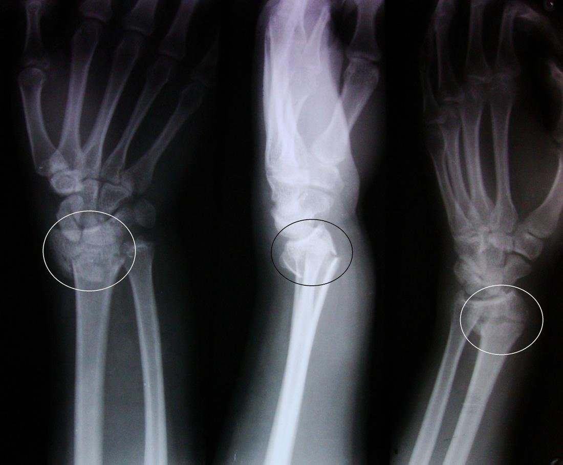
The injury that Abraham Colles described in 1814 is a transverse fracture of the radius just above the wrist, with dorsal displacement of the distal fragments. This injury is caused by a fall on the palm of outstretched hand with supination force. Fracture line lies 2 cm proximal to the distal articular surface of the radius.
Colles’ fracture: Fracture of the lower end of the radius one inch above the lower articular surface of the radius is called colles fracture.
[Ref-Dr. Jahir, “Surgery 1″ Paper” 4th Edition, Page-552]
Or
Colles’ fracture is not just fracture lower end of radius but a fracture dislocation of the inferior radioulnar joint.
[Ref-John Ebnezar’s “Textbook of Orthopedics” 4th edition page-643]
Or
A transverse fracture of the radius just above the wrist, with dorsal displacement of the distal figment is called colles’ fracture.
[Ref-Apley’s “System of Orthopaedics and Fractures” 9th edition page-772]
Clinical features of colics’ fractures :
A) Patients profile: Older age, especially postmenopausal women.
B) Symptoms:
1) History of fall in out stretched hand.
2) Pain and swelling around the wrist and dorsum of the hands.
3) Patients is unable to catch anything.
4) Painful and restricted movement of fingers and wrist.
C) Signs:
1) Dinner fork deformity of the wrist.
2) Hand is radially deviated.
3) Tenderness over the fracture site.
4) Movement of wrist and fingers are restricted.
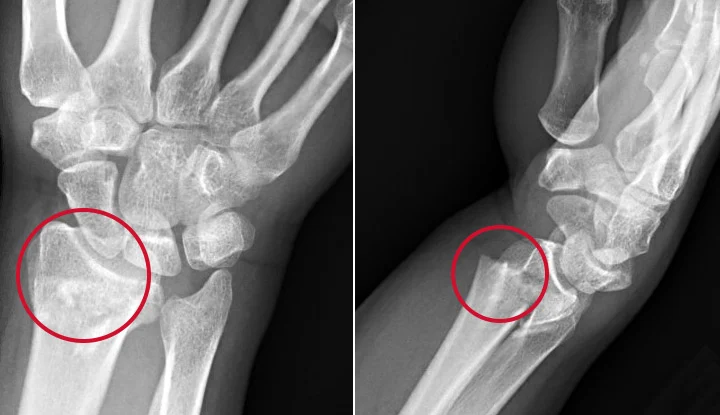
Radiological features of colles fracture: The five commonly seen deformities are:
1. Dorsal angulation with loss of the normal (5-10 degrees) volar tilt of the articular surface of the radius.
2. Dorsal displacement of the distal fracture fragment.
3. Impaction at the fracture site.
4. Radial displacement of the distal fragment.
5. Radial tilt of the distal fragment.
Mechanism:
The common mode of injury is fall on an outstretched hand with dorsiflexion ranging from 40-90° (Average 60°). Displacement: There are six classical displacements in a colles’ fracture:
1. Dorsal displacement.
2. Dorsal rotation.
3. Lateral displacement.
4. Lateral rotation.
5. Impaction.
6. Supination.
(Ref-John Ebnezar’s “Textbook of Orthopedics” 4th edition, page-643,644)
Complications of colics’ fracture:
A) Early complications:
1) Unstable reduction.
2) Median or ulnar nerve stretched.
3) Post reduction-swelling.
4) Compartmental syndrome.
5) Anesthesia problems.
6) Injury to proximal segment of the bone duration reduction.
Radiological features of colles fracture: The five commonly seen deformities are:
1. Dorsal angulation with loss of the normal (5-10 degrees) volar tilt of the articular surface of the radius.
2. Dorsal displacement of the distal fracture fragment.
3. Impaction at the fracture site.
4. Radial displacement of the distal fragment.
5. Radial tilt of the distal fragment.
B) Late complications:
1. Malunion. And nonunion.
2. Rupture of extensor pollicis tendon.
3. Sudeck’s osteodystrophy.
4. Frozen shoulder.
5. Carpal tunnel syndrome.
6. Osteoarthritis of wrist joint.
7. Hand shoulder syndrome.
8. Instability of inferior radioulnar joint.
[Ref-John Ebnezar’s “Textbook of Orthopedics” 4th edition,page-644]
Causes are responsible for malunion of colics’ fracture:
Malunion is the most common complication of Colles’ fracture. Six important causes are responsible for it.
1) Improper reduction: If the fracture is not reduced properly, in the initial stages it may result in malunion later.
2) Improper and inadequate immobilization: This fracture needs to be immobilized at least for a period of six weeks failing which malunion results.
3) Comminuted dorsal surface: Due to extensive comminution, the fracture collapses and recurs after reduction and casting.
4) Osteoporosis may lead to collapse and recurrence.
5) Recurrence: This is due to extensive comminution and osteoporosis.
6) Rupture of the distal radioulnar ligament: This usually goes undetected in the initial stages of treatment and is responsible for the later recurrence.
[Ref-John Ebnezar’s “Textbook of Orthopedics” 4th edition page-652]
There are six options of treatment in a malunited Colles’ fracture:
1. No treatment is required if the patient has no functional abnormality. Immobilization by short arm back slab extending from below elboe to distal metacarpal back slab is replaced by colles full plaster after 24 hours for 4-6 hours.
2. Remanipulation is attempted if fracture is less than 2 weeks old.
3. Darrach’s operation is more often indicated if the patient complains of functional disability.
4. Corrective osteotomy and grafting if the patient wants cosmetic correction and if the patient is young (Fernandez and Campbell).
5. Arthrodesis (for intra-articular fracture): The patient complains of pain in the wrist joint due to traumatic osteoarthritis following an intra-articular fracture. In these patients, arthrodesis of the wrist in functional position is the surgery of choice.
6. Combination of these like Darrach’s operation with osteotomy, etc. is also tried in some situations. Note: Fernandez is a dorsal wedge osteotomy and Campbell is a lateral wedge osteotomy.
[Ref-John Ebnezar’s “Textbook of Orthopedics” 4th edition, page-652]