Concept about Uterus – The course is designed for the basic understanding of anatomical structures and physiological functions of human body, musculoskeletal system, digestive system, respiratory system; cardiovascular system; urinary system, endocrine system, reproductive system, nervous system, hematologic system, sensory organs, integumentary system, and immune system.The aim of the course is to acquire knowledge and skills regarding anatomy and physiology.
Concept about Uterus
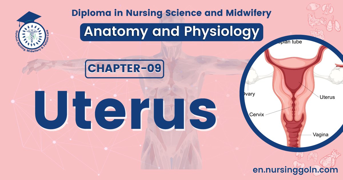
Uterus
The uterus (Also known as womb) is a hollow, muscular, pear-shaped female reproductive organ located posterior and superior to the urinary bladder in the pelvic cavity. Connected to the two fallopian tubes on its superior end and to the vagina (via the cervix) on its inferior end, the uterus is also known as the womb, as it surrounds and supports the developing fetus during pregnancy.
Measurement-
- Length: About 7.5 cm (3 inches).
- Width: About 5 cm (2 inches).
- Thickness: About 2.5 cm (1 inch).
- Weight: About 30-60 gm.
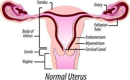
Layers of the wall of uterus :-
There are three layers of uterus wall.
1. Perimetrium (outer layer).
2. Myometrium (middle layer)
3. Endometrium (inner layer): It is a specialized form of mucous membrane with large numbers of mucous-secreting glands which varies in thickness according to the phases of the menstrual cycle.
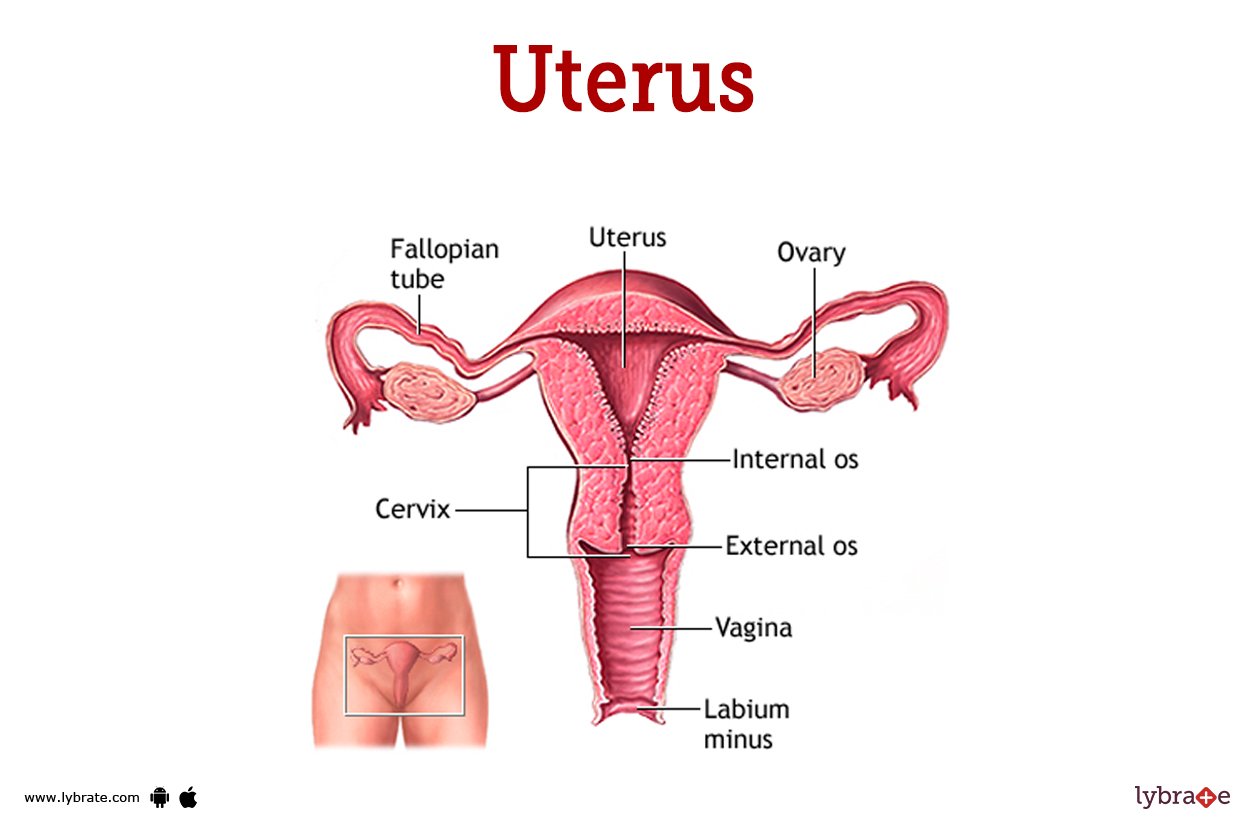
Parts of uterus :-
There are three parts of uterus.
1. The fundus: This is the dome-shaped part of the uterus above the openings of the uterine tubes.
2. The body: It is the main part and forms 2/3 of the organ which extends from the fundus to the internal os of the cervix, where implantation normally occurs.
3. The cervix: The lower narrow part of the uterus is called the cervix. The lumen of the cervix communicates with the cavity of the body of uterus, via internal os (os = mouth) and with that of the vagina via the external os.
Supports of the uterus:
The uterus is supported in the pelvic cavity by surrounding organs, muscles & bones of the pelvic floor and some ligaments that suspended it from the walls of pelvic. Supporting structures are..
- The broad ligaments
- The round ligaments
- The uterosacral ligaments
- The transverse cervical ligaments
- The pubocervical fascia
Blood supply
The uterus is supplied by arterial blood both from the uterine artery and the ovarian artery. Another anastomotic branch may also supply the uterus from anastomosis of these two arteries.
Artery: Ovarian artery and uterine artery
Vein: Uterine veins
Lymphatic drainage
Three main routes constitute the uterine lymphatic drainage. The lymphatic vessels of the uterine fundus mainly drain, like the ovarian vessels, to the aortic or lumbar nodes. The external iliac lymph nodes drain the lymphatics of the uterine body and, occasionally, some lymphatics of the uterine fundus.
Functions of the uterus
1. The endometrium of the uterus undergoes changes in order that it may be prepared to receive a fertilized ovum for implantation.
2. After implantation of fertilized ovum, the uterus develops and enlarges in order to retain and
nourish the developing embryo and fetus.
3. At the end of the pregnancy, the muscular walls of uterus contracts rhythmically and expels the baby and placenta.
4. After expulsion of baby and placenta from uterus, then returns to approximately its normal size by a process known as involution.
Shortly :-
1. The endometrium of the uterus prepared to receive a fertilized ovum for implantation.
2. After implantation, it nourishes the developing embryo and fetus.
3. Expulsion of the baby and placenta from uterus by rhythmic contraction.
(Ref:- R.S. Winwood, Sear’s Anatomy, 6th ed, P-282,283 + G. John, 2nd ed, P-283-285 + P.Evelyn, 16,P-308 310+ Ross & Wilson, 9th, P-441,442)