Concept of Lymphoma – This course is designed to understand the concept of community health nursing: nurses’ roles and interventions in family health, school health, occupational health, environmental health, elderly health care, gender issues, disaster management and principles and terminology of epidemiology. The aim of the course is to acquire knowledge and skills in community health nursing.
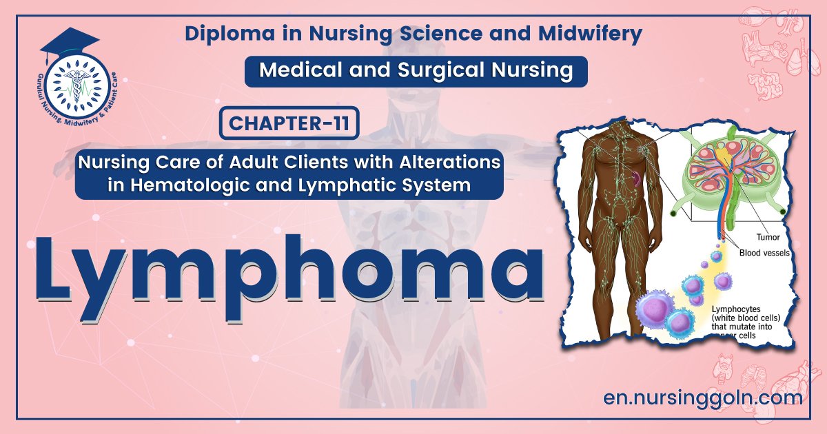
Concept of Lymphoma
Definition of Lymphoma:
Lymphoma is a group of disorders due to neoplastic proliferation of cells of lymphoid series that gives rise to solid tumors of lymphnode structures.
(Ref. de Gruchy’s278/6th)
The lymphatic structures are defined as the lymphh nodes, spleen, thymus, Waldeyer’s ring, appendand Peyer’s patches.
In short; Neoplastic proliferation of lymphoid tissue & majority of B cell origin.
Classification of Lymphoma:
a. Hodgkin disease: Malignant neoplasm of lymphnode with presence of RS (Reed- Sternberg) cells and pleomorphic infiltration by lymphocyte, eosinophil, macrophage, plasma cell, neutrophil &.follicular dendritic cell.
➤ Spread: It is contiguous from one node to the next and extranodal disease, such as bone, brain or skin. Involvement.
➤ Pelebstein fever in Hodgkin disease: Pyrexia period followed by period of apyrexia
➤ Hallmark of Hodgkin’s disease; Reed-Sternberg cells.
Criteria of Reed-Sternberg cells:
- It is a malignant giant cell of B cell origin.
- Large cells with paired mirror-image nuclei.
- This nuclei resembles ‘owl’s eye’ appearance.
- Prominent nucleoli.
b. Non-Hodgkin’s Lymphoma; Malignant neoplasm of lymphnode characterized by monomorphic infiltration and absence of RS (Reed-Sternberg) cells
Histopathological types of classical HL: (Rye classification)
i. Lymphocyte predominant (5%): Prognosis is good
ii. Nodular sclerosing: (70%) Prognosis is good
iii. Mixed cellularity: (20%) Prognosis is not good
iv. Lymphocyte depleted (Rare): Prognosis is not good
The WHO classification Hodgkin disease is based on histology:
i. Nodular lymphocyte- predominant HL. -5%
ii. Classical HL (from the appearance of the Reed-Sternberg cells and surrounding reactive cells):
- Nodular sclerosing 70%
- Mixed cellularity 20%
- A Lymphocyte-rich 5%
- Lymphocyte-depleted (Rare)
Staging of Hodgkin lymphomas:
➤ Stage-1: Involvement of a single lymph node region or extra lymphatic site.
➤ Stage -2: Involvement of two or more lymph node regions or an extra lymphatic site and lymph node regions on the same side of (above or below) the diaphragm.
➤ Stage -3: Involvement of lymph node regions on both sides of the diaphragm with or without localized extra lymphatic involvement or involvement of the spleen or both.
➤ Stage-4: Diffuse involvement of one or more extra lymphatic tissues, e.g. liver or bone marrow
All stages are further divided on the basis of the absence (A) or presence (B) of the following symptoms –
- Unexplained fever
- Drenching night sweats
- Unexplained weight loss of greater than 10% of normal body weight
A symptoms: No systemic symptoms
B-Systemic symptoms: Unexplained Weight loss, drenching sweats, Unexplained fever.

Management of Hodgkin’s disease:
Clinical features:
Age:
- Median age 31 yrs.
- It occurs in adolescence & young adults (20 to 35 yrs of age) & second at 50 to 70 yrs
Sex:
- Sex ratio: 1.5:1
History:
- Past history of infectious mononucleosis (3 times more common)
- More common in patient from well-educated background & small families.
Patient may asymptomatic
i. Lymphadenopathy:
- Usually in the cervical & supraclavicular or mediastinal lymphnode.
- Painless, discrete & rubbery lymphadenopathy & the lymph may fluctuate in size.
ii. B symptoms /Systemic symptoms:
- Fatigue, malaise weight loss, drenching sweats,
- Fever (Pel-ebstein fever), anorexia, pruritus.
iii. Feature of large mediastinal obstruction:
- Dry cough & breathlessness.
- Venous obstruction & dysphasia
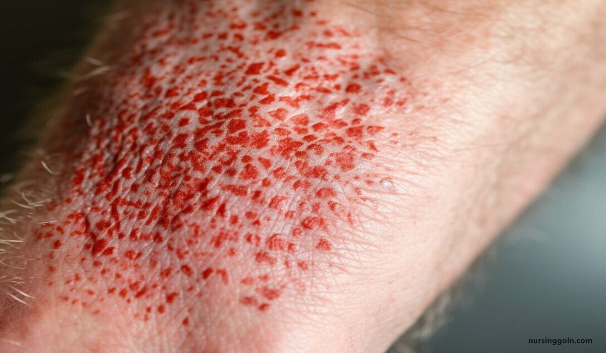
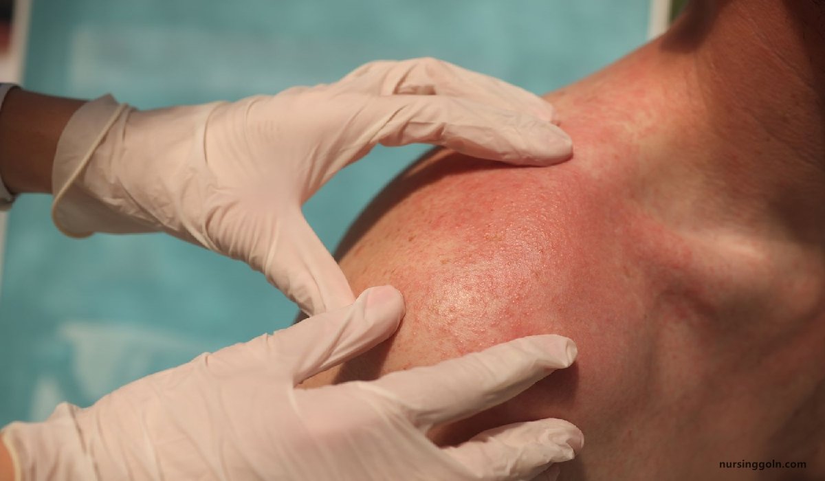
iv. Skin involvement:
- Pruritus, pigmentation & purpura.
v. Abdominal involvement:
- Splenic discomfort, abdominal pain or disturbance of bowel habit.o
vi. Brain involvement:
- Paraplegia, headache, neck stiffness, raised intracranial pressure, weakness.
vii. Bone involvement:
- Bone pain, Anaemia, bleeding manifestation.
ix. Infiltration of viscera:
- Hepatosplenomegaly.
- Pleura or pericardial effusion.
- CCF, pericarditis & arrythmia.
x. Metabolic disturbance:-Hyperuricaemia.
- Gout.
- Renal colic.
- Renal failure.
(Ref. de Gruchy’s 282/6)
Investigations:
i. Full blood count:
- A normochromic normocytic anaemia
- Lymphopenia: Bad prognostic factor.
- An eosinophilia or a neutrophilia
- ESR -raised
ii. For staging & before therapy:
- Renal function tests
- Liver function tests
- LDH measurements, as raised levels are an adverse prognostic factor.
- Chest X-ray may show a mediastinal mass.
- CT scan of chest and abdomen:
✔ Bulky diseases-> 10cm in single node mass -Adverse prognostic factor
- USG of abdomen.
iii. Lymph node biopsy – Confirmity.
iv. Bone marrow study-Shows involvement in patient with advanced diseases
Treatment
i. Radiotherapy:
Indication:
- Stage I disease
- Stage IIA disease with 3 or fewer areas involvement.
- After chemotherapy to sites where there was originally bulk diseases.
- To lesion causing pressure problem.
ii. Chemotherapy:
Indication:
- All pts with B symptoms (Fever, drenching night sweats, wt. loss >10%)
- Stage IIA disease with >3 areas involvement.
- Stage III & IV diseases.
- If relapse occurs after chemotherapy.
Agents & duration: Every 3-4 weeks for total 6-8 cycles.
a. ABVD therapy:
- Adriamycin
- Bleomycin
- Vinblastine
- Dacarbazine.
b. ChIVPP therapy.
- Chlorambucil
- Vincristrine
- Prednisolone
- Procarbazine.
c. COPP therapy.
- Cyclophosphamide,
- Vincristrine
- Prednisolone
- Procarbazine.
iii. Combined modality therapy:
- Radiotherapy given to original site of bulk diseases.
- Rx by chemotherapy to reduce the risk of relapse.
Non-Hodgkin’s Lymphoma
Definition:
Monoclonal proliferation of lymphoid cells of B cell origin (70%) or T cell (30%)
Classification:
WHO classification:
i. Low grade:
- Low proliferation rate
- Asymptomatic for many months before presentation.
- Runs an indolent course
- Is not curable by conventional therapy.
ii. High grade:
- High proliferation rate
- Rapidly produces symptoms, is fatal if untreated.
- It is potentially curable.
Working classification (1982):
i. Low grade:
- Small Lymphocyte
- Folicullar small cleaved cells
- Folicullar mixed cleaved cells & large cells
ii. Intermediate grade:
- Folicular large cell (may be cleft or non-cleft)
- Diffuse small cleaved cell
- Diffuse mixed cleaved cells & large cells
- Diffuse large cell
iii. High grade:
- Immunoblastic cells
- Lymhoblastic
- Noncleaved Burkitt’s lymphoma (CNS involvement- meninges.)
Etiology:
a. Age: 65-70 yrs
b. Sex: slight male excess
c. Virus: HIV, EBV, HSV-8, HTLV,
d. Bacteria: H. pylori. (Gastric lymphoma)
Clinical feature of Non-Hodgkin’s Lymphoma:
1. Age: Median age 65-70 yrs
2. Sex: slight male excess
3. Painless, non-contiguous, mainly peripheral lymphadenopathy (axillary, epitrochlear, mesenteric lymphnode)
4. Systemic upset: fever, sweats, wt. loss, pruritus.
5. Hepatosplenomegaly
6. Extranodal involvement: common.
- BM-Thrombocytopenia & neutropenia, bone marrow failure.
- GIT- Abdominal mass, abdominal distension, intestinal obstruction.
- Thyroid & testis involvement
- Skin-Pruritus, pigmentation & purpura
- Brain-paraplegia
7. Compression syndrome:
- Gut obstruction,
- SVO
- Ascites,
- Spinal cord compression
Investigations:
1. Full blood count:
- A normochromic normocytic anaemia
- Lymphopenia: bad prognostic factor.
- An eosinophilia or a neutrophilia
- ESR-raised.
ii. For staging & before therapy:
- Renal function tests
- Liver function tests
- LDH measurements, as raised levels are an adverse prognostic factor.
- Chest X-ray may show a mediastinal mass.
- CT scan of chest and abdomen.
- USG of abdomen.
iii. Lymph node biopsy – Conformity.
iv. Routine bone marrow aspiration and trephine.
v. Immunophenotyping of surface antigens to distinguish T- and B-cell tumours.
vi. Immunoglobulin determination: For treatment response
vii . Measurement of uric acid levels: High rate can precipitate renal failure.
viii. HIV testing. This may be appropriate if risk factors are present.
Treatment:
I. Low grade NHL:
1. Asymptomatic patients may not require therapy
2. Indications for treatment:
- Marked systematic symptoms.
- Lymphadenopathy causing discomfort or disfigurement.
- Bone marrow failure or compression syndromes.
The options are:
a. Radiotherapy: Stage I disease.
b. Chemotherapy:
- Mainstay of therapy.
- Oral therapy with chlorambucil
c. Monoclonal antibody therapy: Rituximab (Anti-CD20)
- Stage III and IV follicular lymphoma.
d. Transplantation: Studies of autologous stem cell transplantation are in progress.
II. High-grade NHL: Patients with high-grade NHL need treatment at initial presentation.
a. Chemotherapy: Mainstay of therapy. (>90% cases need IV combination therapy) CHOP therapy.
- Cyclophosphamide.
- Doxorubicin
- Vincristrine
- Prednisolone
b. Radiotherapy: Stage I disease patients without bulky disease.
c. Monoclonal antibody therapy: Rituximab combined with CHOP chemotherapy.
d. Transplantation: Autologous stem cell transplantation.
Clinical differences between Hodgkin’s & non-Hodgkin’s lymphoma:
| Traits | Hodgkin lymphoma | Non-Hodgkin Lymphoma |
| Age | Age: 20-35 or 50-70 ута | 65-70 yrs. |
| Lymph node involvement | More often localized to a single axial group of nodes (cervical, mediastinal, para-aortic) | More frequent involvement of multiple peripheral nodes. |
| Lymph node spread | Orderly spread by contiguity | Noncontiguous spread. |
| Mesenteric nodes & Waldeyer ring | Mesenteric nodes & Waldeyer ring rarely involved. | Mesenteric & Waldeyer ring nodes commonly involved. |
| Extranodal involvement | Extranodal involvement uncommon | Extranodal involvement common- liver, spleen, kidney. |
| Systemic features | Systemic features common | Uncommon |
| Pel-Ebstein fever | Pel-Ebstein fever may occur | Pel-Ebstein fever may not occur. |
| Prognosis | High cure rate | Low cure rate (Low grate incurable) |