Deep Vein Thrombosis – An orthopedic nurse is a nurse who specializes in treating patients with bone, limb, or musculoskeletal disorders. Nonetheless, because orthopedics and trauma typically follow one another, head injuries and infected wounds are frequently treated by orthopedic nurses.
Ensuring that patients receive the proper pre-and post-operative care following surgery is the responsibility of an orthopedic nurse. They play a critical role in the effort to return patients to baseline before admission. Early detection of complications following surgery, including sepsis, compartment syndrome, and site infections, falls under the purview of orthopedic nurses.
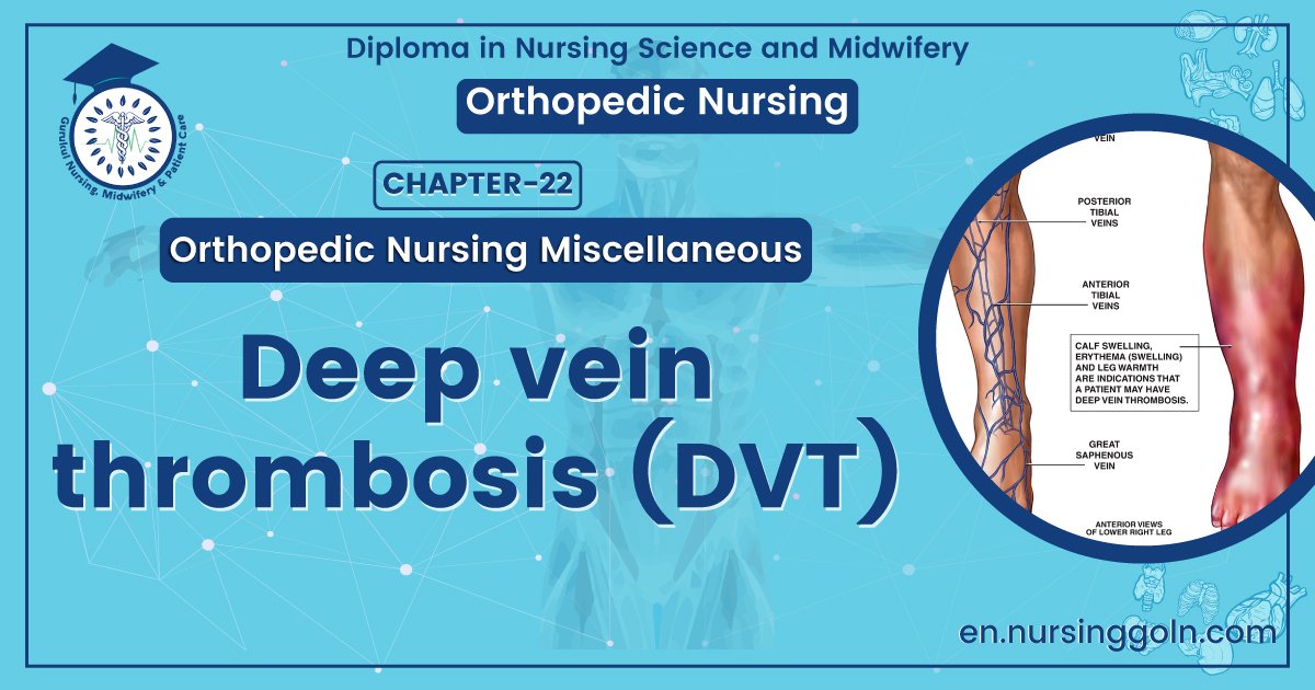
Deep Vein Thrombosis
It is a form of thrombus formation in the deep vein due to complication of fracture of spine, pelvis, femur and tibia.
[Ref-S.das, Clinical surgery, 5th edition, Page-61]
Or
It is a formation of semi-solid(thrombus) in a deep vein. It is a form of thrombophlebitis (inflammation of a vein with clot formation).
[Ref-Baily & Loves, Short Practice of Surgery, 25th edition, Page- 935]
Predisposing factors for development of DVT:
A) Patient factor:
1. Age (>60 years).
2. Obesity.
3. Varicose veins.
4. Immobility.
5. Pregnancy.
6. Puerperium.
7. High dose oestrogen therapy.
8. Previous deep vein thrombosis or pulmonary embolisms.
9. Thrombophilia.
10. Deficiency of antithrombin-III, protein c.
11. Anti-phospholipid.
B) Diseases or Surgical procedure:
1. Surgery of pelvis,hip, lower limb.
2. Malignancy,specially pelvic, abdominal metastatic.
3. Heart failure.
4. Recent myocardial infarction.
5. Paralysis os lower limb.
6. Infection.
7. Inflammatory bowel diseases.
8. Nephrotic syndrome.
9. Paraproteinaemia
10. Homocystinaemia.
[Ref-Baily & loves/25/936]
Common sites of DVT:
1) Pelvic veins-common.
2) Leg veins-femoral vein, popliteal veins.
3) Upper limb veins-Axillary vein.
Clinical features of DVT:
A) Symptoms:
1) Severe calf pain.
2) Swelling & cramp of calf and thigh muscle.
3) Difficulty in standing or walking
4) Pain increase when move or flex leg.

B) Signs:
clinically well detected,
1) Unilateral leg swelling.
2) Increased temperature.
3) Leg is tender, warm pale or bluish and shiny skin.
4) Enlarged superficial veins.
5) Pitting edema.
6) Palbable cord along involved veins.
7) Homans signs +ve
8) Moses signs +ve
(Ref John Ebnezar 44 Surgery for nurses/144)
Investigation confirm the diagnosis:
1. Venous Doppler.
2. Duplex scanning.
3. Ascending Venogram.
4. Others: BT, CT, Prothrombine time, Blood grouping
[Ref-Baily & Loves/25937]
Medical management of DVT:
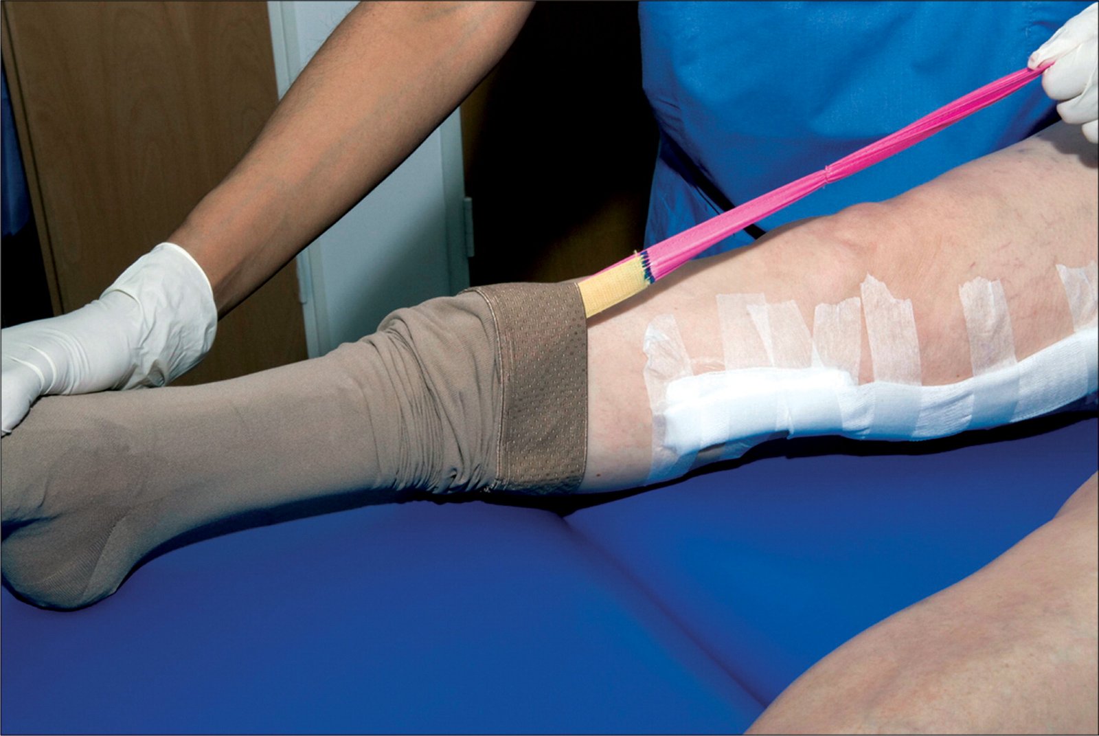
1) Rest: Complete bed rest, elevation of the limb, increase compression whole limb by DVT socking or crape bandage
2) Anticoagulant therapy: Heparine, warfarin, phenindione.
3) Thrombolytic:
a) For fixed thrombus: Initially high dose of heparin of 25,000 units/ day for 7 days Later pts is advised to continue warfarin for 6 months.
b) For free thrombus: Streptokinese (6 lakh units) stat then 1 lakh hourly.
4) Surgery: venous thrombectomy when above measure is not recover or multiple venous thrombosis at a veins.
[Ref Baily & loves/25 1937+SRBS, Surgery for nurses-144]
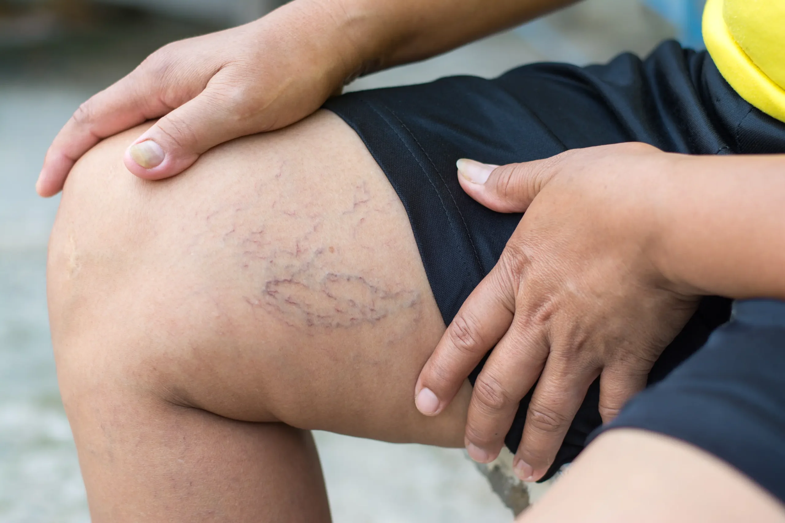
Complication of DVT:
1. Pulmonary embolism
2. Localized edema
3. Venous ulcer
4. Venous gangrene
5. Varicose vein
6. Calcification of vein
7. Severe calf pain.
8. PUO(pyrexia of unknown origin)
9. Chronic venous hypertension
10. Recurrent of DVT
11. Pigmentation.

Differential diagnoses of DVT:
1) DVT.
2) Calf muscle haematoma.
3) Skin inflammation including cellulites.
4) Superficial thrombophlebitis.
Prevent arising complication:
a. Possible diagnosis: DVT.
b. Cardinal signs:
1. Severe calf pain increase on movement.
2. Superficial vein swelling.
3. Homans signs =+ ve.
c. Prevention of complication of DVT:
1) General measures:
a) Early detection of risk factors
b) Proper positioning of leg with no pressure on calf muscle.
c) Elevation of the affected limb.
d) Pressure bandage applied to the leg after major surgery.
e) Stop smoking which increases viscosity of blood
f) Stop OCP before 6-8 weeks of surgery (female)
g) Active or passive exercise of leg

2) Physical measures : To prevent sluggish flow of blood
a) Graduated compression stocking: stocking below knee may be effective than above knee.
b) Intermittent planter vessous compression: intermittent foot pump can increase circulation on the venous plexus.
c) Intermittent pneumatic compression of leg. this compression is useful in Hip replacement.
d) Inferior venacaval filters: Embolus is filters in the inferior venacava before reaching lungs.
e) Electrical stimulation of calf muscle
3) Chemical methods:
a) Aspirin
b) 500 nd of Destrevenously during surgery and another 500 ml in 1 24 boses of postoperative period
c)Heparins 5000 units gives hours before surgery and another 5.000 units continued for 5 days Beaty
Homan’s signs:
- It is a clinical signs of DVT which is examined in calf muscle.
- Passive forceful dorsiflexion of the foot with the knee extended will elicit tenderness in the calf muscle.
- When forced ankle dorsiflexion produce calf pain Homan’s signs are positive.
- When Homan’s sign is positive is significant of DVT.
Moses signs:
Squeezing of relaxed calf muscle from side to side causes pain in the calf muscle. Positive in DVT.
Read more: