
Definition of Pulse – Nursing is a profession within the healthcare sector focused on the care of individuals, families, and communities so they may attain, maintain, or recover optimal health and quality of life. Nurses may be differentiated from other healthcare providers by their approach to patient care, training, and scope of practice. Nurses practice in many specialisms with differing levels of prescriber authority.
Many nurses provide care within the ordering scope of physicians, and this traditional role has shaped the public image of nurses as care providers. However, nurses are permitted by most jurisdictions to practice independently in a variety of settings depending on training level. In the postwar period, nurse education has undergone a process of diversification towards advanced and specialized credentials, and many of the traditional regulations and provider roles are changing.
Nurses develop a plan of care, working collaboratively with physicians, therapists, the patient, the patient’s family, and other team members, that focus on treating illness to improve quality of life. Nurses may help coordinate the patient care performed by other members of an interdisciplinary healthcare team such as therapists, medical practitioners, and dietitians. Nurses provide care both interdependently, for example, with physicians, and independently as nursing professionals.
Definition of Pulse
It is the rhythmic expansion & elongation of the arterial wall passively produced by the pressure changes during ventricular systole & diastole. Range: 60-90/ min (average: 72/min).
Or
Pulse is the rhythmic dilatation and elongation of arterial wall as a result of pressure changes created by the intermittent ejection of blood from heat to the already full aorta feeding the arterial system; transmit as a wave to the periphery.
Or,
The rhythmic dilation of an artery that results from beating of the heart. Pulse is often measured by feeling the arteries of the wrist or neck
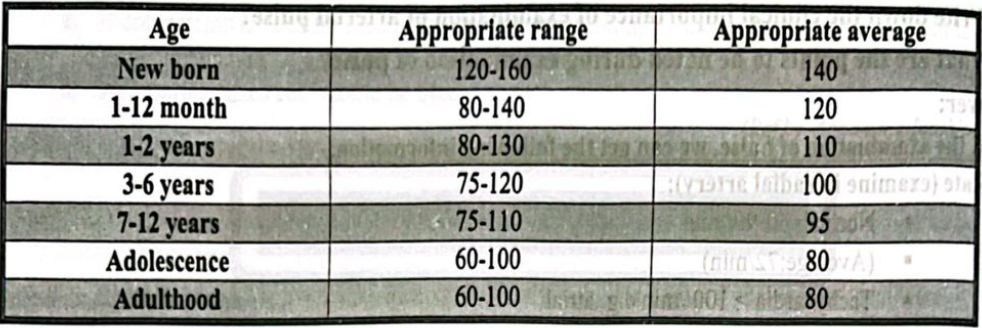
Normal Human Pulse Rate in Human Body:
➤ Range: 60-90/ min
➤ Average: 72/min
Types of Pulse:
A. Arterial pulse
a) Normal or catacrotic
b) Abnormal pulse
c) Anacrotic pulse
d) Dicrotic pulse
e) Collapsing pulse
f) Pulses paradoxes
g) Pulsus patterns
h) Pulsus deficit
B. Venous pulse
From the examination of pulse, we can get the following information:
A. Rate (examine in radial artery):
- Normal: 60-90/ min
- (Average: 72/min)
- Tachycardia > 100/min e.g. atrial fibrillation.
- Bradycardia <60/min. e.g. Complete heart block.
B. Rhythm (examine in radial artery):
C. Character (examine in carotid artery):
- Normal: Catachrestic pulse.
- (Abnormal pulse)
D. Volume (examine in carotid artery):
a) Small volume pulse:
- Shock,
- Aortic stenosis
- Pericardial effusion
b) High volume pulse:
- Hyperdynamic circulation
- Aortic regurtitation
E. Symmetry-
- Compare radial artery of both side.
- Bilateral symmetrical / Radio- radial delay
F. Condition of the vessel wall- in radial artery
- e.g. Just palpable Normal
- Cord like structure (thickened) Atheroslerosis.
G. Any delay between radial & femoral pulse.
- e.g. coaraction of aorta
Normal Range of Pulse Rate:
Factors affecting Pulse Rate:
A. Air temperature: When temperatures (and the humidity) soar, the heart pumps a little more blood, so your pulse rate may increase, but usually no more than five to 10 beats a minute.
B. Body position: Resting, sitting or standing, your pulse is usually the same. Sometimes as you stand for the first 15 to 20 seconds, your pulse may go up a little bit, but after a couple of minutes it should settle down. Emotions: If you’re stressed, anxious or “extraordinarily happy or sad” your emotions can raise your pulse.
C. Body size: Body size usually doesn’t change pulse. If you’re very obese, you might see a higher resting pulse than normal, but usually not more than 100.
D. Medication use: Meds that block your adrenaline (beta blockers) tend to slow your pulse, while too much thyroid medication or too high of a dosage will raise it.
Common Sites of Measuring Pulse:
A. Upper limb
a) Axillary pulse: located inferiorly of the lateral wall of the axilla
b) Brachial pulse: located on the inside of the upper arm near the elbow, frequently used in place of carotid pulse in infants (brachial artery)
c) Radial pulse: located on the lateral of the wrist (radial artery). It can also be found in the anatomical snuff box.
d) Ulnar pulse: located on the medial of the wrist (ulnar artery).
B. Lower limb
a) Femoral pulse: located in the inner thigh, at the mid-inguinal point, halfway between the pubic symphysis and anterior superior iliac spine (femoral artery)
b) Popliteal pulse: Above the knee in the popliteal fossa, found by holding the bent knee. The patient bends the knee at approximately 124°, and the physician holds it in both hands to find the popliteal artery in the pit behind the knee (Popliteal artery).
c) Dorsalis pedis pulse: located on top of the foot, immediately lateral to the extensor of hallucis longus (dorsalis pedis artery).
d) Tibialis posterior pulse: located on the medial side of the ankle, 2 cm inferior and 2 cm posterior to the medial malleolus (posterior tibial artery). It is easily palpable over Pimenta’s Point.
C. Head and neck
a) Carotid pulse: located in the neck (carotid artery). The carotid artery should be palpated gently and while the patient is sitting or lying down. Stimulating its baroreceptors with low palpitation can provoke severe bradycardia or even stop the heart in some sensitive persons.
Also, a person’s two carotid arteries should not be palpated at the same time. Doing so may limit the flow of blood to the head, possibly leading to fainting or brain ischemia. It can be felt between the anterior border of the sternocleidomastoid muscle, above the hyoid bone and lateral to the thyroid cartilage.
b) Facial pulse: located on the mandible (lower jawbone) on a line with the corners of the mouth (facial artery).
c) Temporal pulse: located on the temple directly in front of the ear (superficial temporal artery).
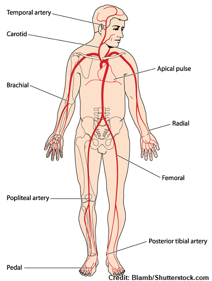
Different common sites of assessing pulse
a) Apical pulse: located in the 5th left intercostal space, 1.25 cm lateral to the mid- clavicular line. In contrast with other pulse sites, the apical pulse site is unilateral, and measured not under an artery, but below the heart itself (more specifically, the apex of the heart). See also apex beat.
Atypical/Abnormal Pulses:
A. Abnormal volume
- Small /Low volume pulse
- High volume pulse
B. Abnormal character:
- Slow rising (anacrotic) pulse
- Collapsing (water-hammer) pulse
- Pulsus bisferiens – ‘double peak
- Pulsus alternans
- Pulsus paradoxus
- Pulsus “deficit”
Causes of an Irregular Pulse
1. Sinus arrhythmia
2. Atrial extrasystoles
3. Ventricular extrasystoles
4. Atrial fibrillation
5. Atrial flutter with variable response
6. Second-degree heart block with variable response
Procedure of Taking Pulse:
1. Wash hands/hand hygiene.
- Rationale: Reduces transmission of microorganisms.
2. Inform client of the site(s) where pulse will be measured.
- Rationale: Encourages participation and allays anxiety.
3. Flex client’s elbow and place lower part of arm across chest. Maintains wrist in full extension and esexposes artery for palpation.
- Rationale: Placing client’s hand over chest will facilitate later respiratory assessment without undue attention to the nurse’s action. (It is difficult for any person to maintain a normal breathing pattern when someone is observing and measuring.)

4. Support client’s wrist by grasping outer aspect with thumb.
- Rationale: Stabilizes wrist and allows for pressure to be exerted.
5. Place index and middle fingers on inner aspect of client’s wrist over the radial artery, and apply light but firm pressure until pulse is palpated. Fingertips are sensitive, facilitating palpation of pulsating pulse. The nurse may feel his or her own pulse if palpating with thumb.
- Rationale: Applying light pressure prevents occlusion of blood flow and pulsation.
6. Identify pulse rhythm. Palpate pulse until rhythm is determined.
- Rationale: Describe as regular or irregular.
7. Determine pulse volume. Quality of pulse strength is an indication of stroke volume.
- Rationale: Describe as normal, weak, strong, or bounding.
8. Count pulse rate by using second hand on watch. For a regular rhythm, count number of beats for 30 seconds and multiply by 2. For an irregular rhythm, count number of beats for a full minute, noting number of irregular beats.
- Rationale: An irregular rhythm requires a full minute of assessment to identify the number of inefficient cardiac contractions that fail to transmit a pulsation, referred to as a skipped or irregular beat.