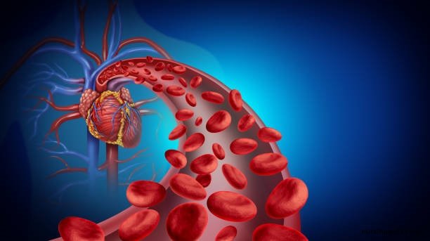
Today our topic of discussion is ” Development of Blood Vessels and Fetal Circulation “. The creation and maturation of the cardiovascular system is one of the earliest and most critical events during embryonic development. The unique nature of fetal circulation, adapted to the intrauterine environment, showcases the dynamic adaptability of the human body. This article delves into the development of blood vessels and the fascinating world of fetal circulation.

Development of Blood Vessels and Fetal Circulation: Blood Vessels and Circulation
1. Introduction
The cardiovascular system’s development begins in the early embryonic stage, evolving through a series of stages that result in a complex network of blood vessels and a functional heart. Simultaneously, the fetus develops a specialized circulation system, distinct from postnatal circulation, to accommodate life within the womb.
2. Angiogenesis and Vasculogenesis
Blood vessel formation during embryonic development is a result of two processes:
- Vasculogenesis: This process refers to the de novo formation of blood vessels. Mesodermal cells differentiate into angioblasts, which then form blood islands. These islands coalesce to form the primitive vascular network.
- Angiogenesis: It involves the sprouting, splitting, and remodeling of existing vessels to form new ones. Endothelial cells play a pivotal role, migrating and proliferating to create new vascular channels.
3. Heart Development
The heart emerges from the mesodermal layer. A straightforward tube forms initially, which then undergoes folding, division, and septation to transform into a four-chambered heart. This intricate process occurs in a sequence of stages:
- Formation of the Cardiac Tube: Two endocardial tubes form and fuse, creating a singular cardiac tube.
- Looping: The cardiac tube undergoes dextral looping, creating the basic shape of the mature heart.
- Septation: Internal walls or septa form, dividing the heart into chambers.

4. Fetal Circulation: An Overview
Given that the fetal lungs are not functional in the womb, fetal circulation bypasses the lungs and relies heavily on the placenta for oxygen and nutrient exchange. This necessitates specialized structures and pathways:
- Placenta: This organ connects the fetus to the maternal uterus, allowing nutrient uptake, waste elimination, and gas exchange via the mother’s blood supply.
- Umbilical Vein: Carries oxygenated blood from the placenta to the fetus.
- Umbilical Arteries: Two arteries that carry deoxygenated blood and waste products from the fetus back to the placenta.
5. Specialized Shunts in Fetal Circulation
Three crucial shunts redirect blood flow in fetal circulation:
- Ductus Venosus: This shunt bypasses the liver, allowing oxygen-rich blood from the umbilical vein to flow into the inferior vena cava.
- Foramen Ovale: An opening in the atrial septum. It allows blood to flow directly from the right atrium to the left atrium, bypassing the non-functioning fetal lungs.
- Ductus Arteriosus: Connects the pulmonary artery to the descending aorta. This ensures that minimal blood reaches the lungs, directing it instead to the body’s vital organs.
6. Transition to Postnatal Circulation
At birth, the physical act of taking the first breath and the sudden exposure to a new environment initiates crucial changes:
- Closure of the Foramen Ovale: The increase in blood returning from the now-functional lungs to the left atrium and the decrease in blood flow from the inferior vena cava causes the foramen ovale flap to close, eventually fusing and becoming the fossa ovalis.
- Closure of the Ductus Arteriosus: Elevated oxygen levels cause this shunt to constrict and eventually close, transforming into the ligamentum arteriosum.
- Closure of the Ductus Venosus: After the umbilical cord’s clamping, this shunt closes and becomes the ligamentum venosum.
7. Clinical Considerations
Occasionally, complications arise in cardiovascular development:
- Patent Foramen Ovale (PFO): In some individuals, the foramen ovale fails to seal completely post-birth, which can lead to health issues in adulthood.
- Patent Ductus Arteriosus (PDA): Failure of the ductus arteriosus to close can result in heart strain and increased pulmonary blood flow.

8. Conclusion
The development of blood vessels and the intricacies of fetal circulation illuminate the marvel of human life. From the earliest stages, the cardiovascular system adapts, grows, and changes to support the body both inside and outside the womb. Understanding these processes reinforces the complex choreography of nature, resulting in the symphony of life that every heartbeat echoes.
