
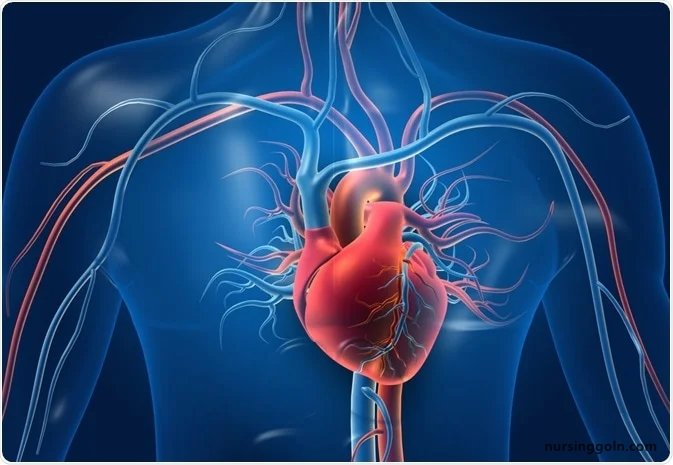
Today our topic of discussion is ” Development of the Heart “. The human heart, a symbol of life, emotion, and vitality, is central to the circulatory system. Its development is a meticulously coordinated process, bringing together cellular differentiation, migration, and organogenesis. This article offers a deep dive into the intricacies of cardiac embryology, chronicling the stages that give rise to this marvelous organ.
Development of the Heart – The Cardiovascular System: The Heart
1. The Setting Stage: Early Embryonic Period
In the initial days post-fertilization, the embryo transforms from a simple zygote to a multi-layered entity. By day 15-20 post-fertilization, three distinct layers are discernible:
- Ectoderm: The outermost layer, giving rise to the nervous system and skin.
- Mesoderm: The middle layer, the precursor for musculature, the skeletal system, and the circulatory system.
- Endoderm: The inner layer, forming the gastrointestinal and respiratory tracts.
The heart’s genesis is deeply intertwined with the mesoderm.
2. Foundation of the Heart Tube
Around the third week, specific regions of the mesoderm termed the splanchnic mesoderm, begin to form paired structures known as angiogenic clusters. These structures coalesce to form two primitive blood vessels, which eventually fuse into a singular, primary heart tube.
3. Dextral Looping
In an almost balletic motion, the heart tube elongates and loops to the right, setting the stage for the differentiation of heart chambers. This dextral looping ensures that the parts of the heart tube develop into their destined structures.
4. Chamber Specification and Division
Post-looping, the heart tube differentiates into five distinct regions:
- Truncus Arteriosus: Develops into the ascending aorta and pulmonary trunk.
- Bulbus Cordis: Transforms into the right ventricle’s smooth parts.
- Primitive Ventricle: Gives rise to the left ventricle.
- Primitive Atrium: Eventually forms the atrial chambers.
- Sinus Venosus: Differentiates into the superior and inferior vena cava and parts of the right atrium.
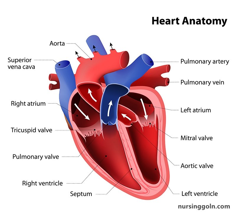
Further, septa form to separate the atria and ventricles, ensuring the heart’s four-chambered design.
5. Formation of the Heart Valves
Valves are critical for the unidirectional flow of blood. Initially, swellings known as endocardial cushions appear in the atrioventricular canal and truncus arteriosus. These cushions proliferate, fuse, and remodel to form the four primary heart valves.
6. Development of the Conductive System
For coordinated contractions, the heart requires an intricate electrical system. Specific clusters of myocardial cells, influenced by underlying genetic factors, differentiate into the SA node, AV node, and the associated conductive pathways.
7. Coronary Circulation Development
The heart, despite being a blood-filled organ, requires its own dedicated blood supply. The coronary arteries emerge from the aorta, growing and branching to enwrap the heart, ensuring its oxygenation.
8. Maturation and the Fetal Circulation
Within the confines of the womb, the fetal heart supports a slightly different circulatory paradigm, necessitating structures like the foramen ovale (a connection between the two atria) and ductus arteriosus (a vessel connecting the pulmonary artery to the aorta). These allow the bypass of the fetal lungs, which are not yet functional.
9. Birth: A Cardiac Revolution
With the first breath, the newborn’s cardiac system undergoes a rapid transformation. The pulmonary resistance drops, the lungs inflate, and structures like the foramen ovale and ductus arteriosus begin to close, establishing the typical postnatal circulation.
10. Genetic and Environmental Influences
Cardiac development is tightly regulated by a plethora of genes. Mutations or dysregulation can result in congenital heart defects. Moreover, maternal factors, including infections, drug use, or uncontrolled diabetes, can impinge on the normal cardiac development of the fetus.
11. Congenital Heart Defects: When Development Goes Awry
Despite the precision of cardiac development, errors can arise. These congenital anomalies, ranging from septal defects to complex structural abnormalities, underscore the importance of meticulous embryonic coordination.
12. The Future: Regenerative Medicine and Cardiac Development
Understanding cardiac embryology holds promise for the burgeoning field of regenerative medicine. By harnessing insights from developmental biology, scientists aim to engineer cardiac tissues, offering hope for heart disease patients.
Conclusion
The heart’s development is a testament to nature’s orchestration prowess. From a simple tube to a four-chambered powerhouse, the journey of the heart encapsulates the miracles of embryology. As we continue to unveil the secrets of cardiac development, we not only deepen our appreciation of this vital organ but also stride toward pioneering therapeutic innovations, reminding us of the wonder of life’s rhythm.