Congenital & Developmental Disorders – An orthopedic nurse is a nurse who specializes in treating patients with bone, limb, or musculoskeletal disorders. Nonetheless, because orthopedics and trauma typically follow one another, head injuries and infected wounds are frequently treated by orthopedic nurses.
Ensuring that patients receive the proper pre-and post-operative care following surgery is the responsibility of an orthopedic nurse. They play a critical role in the effort to return patients to baseline before admission. Early detection of complications following surgery, including sepsis, compartment syndrome, and site infections, falls under the purview of orthopedic nurses.
Congenital & Developmental Disorders
Definition of congenital disorders:
Congenital disorders are defined as those abnormalities of development that are present at the time of birth. [Ref-John Ebnezar’s, “Textbook of Orthopedics”, 4″ edition, P-487)
Or
The term congenital anomaly and congenital malformation are used interchangeably, but they are different in meaning. A WHO document described the terms, the congenital anomaly being used to include all biochemical, structural and functional disorders present at birth and the congenital malformation should be confined to structural defects only, present at birth. The birth defects or congenital defects may be inherited generally or acquired during gestation or inflicted during parturition.
Common congenital disorders:
1. Generalized congenital abnormalities:
i. Achondroplasia.
ii. Diaphysial Aclasis.
iii. Dyschondroplasia (Oilier’s Disease),
iv. Cranio-cleidal Dysostosis.
v. Osteogenesis Imperfecta.
vi. Arthrogryposis Multiplex Congenita.
2. Local congenital abnormalities:
i. Spina Bifida.
ii. Congenital Vertical Talus. Abnormalities of Digits
3. Congenital disorders of trunk and upper extremity:
i. Congenital torticollis.
ii. Congenital elevation of scapula,
iii. Congenital pseudarthrosis of the clavicle,
iv. Congenital radioulnar synostosis.
4. Congenital disorders of hip and pelvis:
i. Congenital dislocation of hip.
ii. Congenital coxa vara.
5. Congenital disorders of the lower limb:
1. Congenital dislocation of knee,
ii. Congenital pseudarthrosis of the tibia
iii. Congenital talipes equinovarus (CTEV) or idiopathic club foot.
6. Congenital absence of part or all of long bones:
i. Radius,
ii. Tibia,
iii. Fibula.
[Ref-J N ASTON’s, “Orthopedics and Traumatology”, 2 edition, P-193 & Ebnezar’s, “Textbook of Orthopedics”, 4″ edition, P-487]
Causes of congenital disorder in BD:
The exact cause is unknown. Most congenital disorder begin early in the life of the embryo when cell division is most active. Although a few congenital disorders may be due to uterine malposition, most are believed to be due genetic defects, environmental influences or a combination of both.
A. Genetic factors:
Congenital anomalies are inherited through the genes in the ovum and sperm. The anomalies may related to:
1. Chromosomal abnormalities
2. Single gene disorderor polygenic inheritance
B. Environmental factors:
Various environmental factors are responsible for occurrence of congenital anomalies. Teratogenesis agents of the environment adversely affects the normal cellular development of the embryo or fetus causing birth defects. The common environmental factors are:
1. Intrauterine infection especially by STORCH.
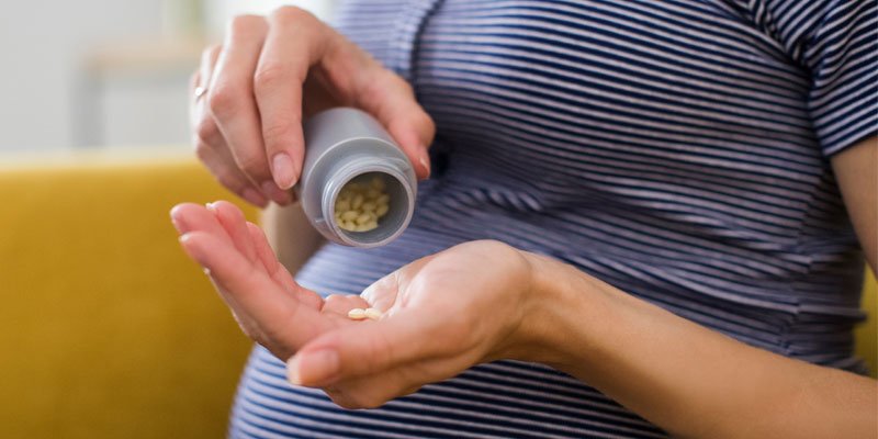
2. Drugs intake by mother during pregnancy like steroid hormons, stilbestrol, anticonvalsant cocaine, lithium etc.
3. X-ray exposure during pregnancy.
4. Meternal diseases like diabetes, cardiac failure, malnutrition, iodine deficiency disorder etc.
5. Abnormal uterine environment like bicornute uterus, septed uterus, fetal hypoxia etc.
6. Maternal addiction with alcohol, tobacco or smoking. 7. Environmental pollution especially air pollution.
C. Embryonic Trauma:
Congenital disorders can also result from injury to the developing embryo at the time of differentiation.
[Ref- Parul Datta’s, “Pediatric Nursing”, 2″ edition, P-196]
Teratogenesis
Definition of teratogenesis: Experimental production of congenital anomalies is teratogenesis.
[Ref-John Ebnezar’s, “Textbook of Orthopedics”, 4 edition, P-488]
Factors of teratogenesis:
1. Metabolic.
2. Hormonal.
3. Nutritional deficiency.
4. Chemicals.
5. X-ray.
6. Trauma.
7. Infection.
8. Mechanical.
9. Thermal.
10. Anoxia etc.
[Ref-John Ebnezar’s, “Textbook of Orthopedics”, 4″ edition, P-487]
Congenital Torticollis (Wryneck)
Definition of Congenital Torticollis:
Congenital Torticollis is a condition where the sternocleidomu muscle of the neck undergoes contractures pulling it to the same side and turning the face of the opposite side.
(Ref-John Ebnezar’s, “Textbook of Orthopedics”, 4″ edition, P-488)
Causes of Congenital Torticollis :
1. The exact cause is unknown.
2. Hypothetically it may due to fibromatosis within the sternomastoid muscle.
[Ref-John Ebnezar’s, “Textbook of Orthopedics”, 4″ edition, P-488]
Clinical presentations of Congenital Torticollis:
1. Tumor palpable at birth or during the first two weeks of life.
2. Common on the right side.
3. May include the muscle diffusely but more often it is localized near the clavicular attachment of the muscle.
4. It attains maximum size within 1-2 months; usually it disappears within a year.
5. If it fails to disappear, then the muscle becomes permanently fibrotic and contracted and cause torticollis.
[Ref-John Ebnezar’s, “Textbook of Orthopedics”, 4″ edition, P-488]
Principles treatment of Congenital Torticollis:
1. During infancy, conservative treatment consists of stretching of the sternomastoid by manipulation and physiotherapy. Excision is unjustified in infancy.
2. Surgery is delayed until fibroma is well formed.
3. If the muscle is still contracted at the age of 1 year, it should be released.
4. If wryneck is persistent for 1 year, it will not resolved spontaneously and needs to be interfered operatively.
5. Exercise program is successful:
i. When restriction of motion is less than 30°.
ii. When there is no facial asymmetry.
6. Non-operative treatment after 1 year is rarely successful.
7. Surgical method: Subcutaneous tenotomy of the clavicular attachment of the sternomastoid muscle.
[Ref-John Ebnezar’s, “Textbook of Orthopedics”, 4″ edition, P-489]