
Diet for a Patient of Burn – This book covers the entire syllabus of “Nutrition and Dietetics” prescribed by BNMC for all Diploma in Nursing Science and Midwifery students. We tried to accommodate the latest information and topics. This book is an examination-friendly setup according to the teachers’ lectures and examination questions.
At the end of the book previous university questions are given. We hope in touch with the book students’ knowledge will be upgraded and flourish. The unique way of presentation may make your reading of the book a pleasurable experience.
Diet for a Patient of Burn
Definition of Burn:
According to WHO (World Health Organization) “A burn is an injury to the skin or other organic tissue primarily caused by heat or due to radiation, radioactivity, electricity, friction or contact with chemicals. Skin injuries due to ultraviolet radiation, radioactivity, electricity or chemicals, as well as respiratory damage resulting from smoke inhalation, are also considered to be burns”

Burns may be defined as injuries resulting from the application of dry heat (e.g. flame and heated solid substances) or chemical substances to the external or internal surfaces of the body resulting in more or less destruction of the tissues.
Or,
Damage to the skin or other body parts caused by extreme heat, flame, contact with heated objects, or chemicals is called burn. Burn depth is generally categorized as first, second, or third degree Burns can be very painful and may cause:
- Red or Peeling Skin
- Blisters
- Swelling
- White or Charred Skin
(Ref by-www.medicinenet.com)
Definition of Scalds:
Scald is a form of thermal burn injury resulted from heated fluids such as boiling water or steam. Most scalds are considered first or second degree burns, but third degree burns can result, especially with prolonged contact.

Scalds may be defined as injuries produced by the application of moist heat (e.g. a liquid at or near its boiling point or in its gaseous form such as steam) to the body.
(Ref by- Bailey & Love’s/24th/270+23rd/189)
Classification of Burn
Burn may be classified as-
I. According to depth of tissue damage
II. According to cause
III. According to zone/extent(rule of nine)
A. According to the depth of tissue damage:
a) Traditional
- First degree: Includes only the outer layer of skin, the epidermis. Skin is usually red and very painful
- Second degree: Second-degree burns involve the epidermis and part of the dermis layer of skin. The burn site appears red, blistered, and may be swollen and painful.
- Third degree: All layers of the skin are destroyed. Extends into the subcutaneous tissues
- Fourth degree: Same as third degree but with damage to deeper structures such as tendons, joints and bon


b) Modern terminology:
- Superficial:- Involves only the outer epidermis; heals without scarring
- Superficial Partial-Thickness: Involves epidermis & the upper portion of the dermis; pain & blisters; heals with min to no scarring
- Deep-Partial Thickness:- Complete destruction of the epidermis & the majority of the dermis; blisters, edema; may heals with hypertrophic scars & keloids
- Full-Thickness: Complete destruction of the epidermis & dermis along with partial damage of the subcutaneous fat layer; require grafts & susceptible to infection
- Sub-dermal: Complete destruction of the epidermis, dermis, & subcutaneous tissue; may involve muscle & bone; often requires surgical intervention
B. According to cause
a) Thermal Burn: Caused by conduction or convection Example. Hot liquid, fire or steam
b) Electrical Burn: Caused by the passage of electrical current through the body. There is typically an entrance & an exit wound.
c) Chemical Burn: Occurs when certain chemical compounds come in contact with the body. Example. Sulfuric acid, lye, hydrochloric acid, gasoline
C. According to zone/extent
a) Zone of Coagulation: Area of greatest destruction, tissue necrosis, irreversible cell damage
b) Zone of Stasis: Damaged tissue, area of less severe injury that possesses reversible damage and surrounds the Zone of Coagulation
c) Zone of Hyperemia: Pink, no cell death, the area surrounding the Zone of Stasis that presents with inflammation, but will fully recover without any intervention or permanent damage.
(Ref by- Bailey & Love’s/24th/270+234/189)
Difference of First, Second and Third Degree Burn
| Traits | First | Second (Superficial or Deep) | Third (Full Thickness) |
| Depth (how deep the burn is) | Epithelium | Epithelium and top aspects of the dermis | Epithelium and dermis |
| How the wound looks | No blisters; dry pink | Moist, oozing blisters; Moist, white, pink, to red | Leathery, dry, no elasticity; charred appearance |
| Causes | Sunburn, scald, flash flame | Scalds, flash burns, chemicals | Contact with flame, hot surface, hot liquids, chemical, electric |
| Level of Pain (sensation) | Painful, tender, and sore | Very painful | Very little pain, or no pain |
| Healing Time | Two to five days; peeling | Superficial: five to 21 days. Deep: 21-35 days | Small areas may take months to heal; large areas need grafting. |
| Scarring | No scarring; may have discoloration | Minimal to no scarring; may have discoloration | Scarring present |
| Burn injury is the destruction of the layers of the skin and associated structures. | ||
 | 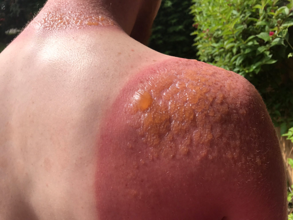 | 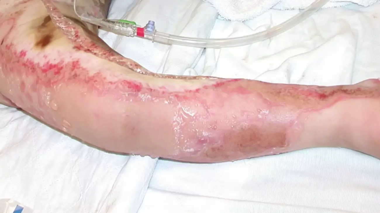 |
| 1st Degree Burn | 2nd Degree Burn | 3rd Degree Burn |
| | |
Causes of Burn:
Many things can cause burns, including:
1. Fire
2. Hot liquid or steam
3. Hot metal, glass or other objects
4. Electrical currents
5. Radiation from X-rays or radiation therapy to treat cancer
6. Sunlight or ultraviolet light from a sunlamp or tanning bed
7. Chemicals such as strong acids, lye, paint thinner or gasoline
8. Flames
9. Lighting
10. Friction
Causes of Scalds:
1. Boiling water
2. Steam from boiling water
3. Any other hot liquid such as tea, coffee, hot oils etc.
Sign and Symptoms of Burn:
1. Blisters
2. Pain (the degree of pain is not related to the severity of the burn — the most serious burns can be painless)
3. Peeling skin
4. Red skin
5. Shock (watch for pale and clammy skin, weakness, bluish lips and fingernails, and a drop in alertness)
6. Swelling
7. White or charred skin
8. Charred mouth; burned lips
9. Burns on the head, face, or neck
10. Wheezing
11. Change in voice
12. Difficulty breathing; coughing
13. Singed nose hairs or eyebrows
14. Dark, carbon-stained mucus
Full thickness burn and sub-dermal burn are so dangerous because-
- Full thickness burn completely destroys the epidermis & dermis along with partial damage of the subcutaneous fat layer.
- Sub-dermal burn completely destroys of the epidermis, dermis, & subcutaneous tissue; may involve muscle & bone.
Burn is dangerous because:
1. Burn injury increases the risk of sepsis which is life threatening infection that rapidly travels through bloodstream.
2. There are lots of complications may arise due to burn on the basis of we can say that burn is so dangerous. The following reasons are given below-
- Burn can damage blood vessel
- Chance of massive infection
- Scarring
- Bone and joint problem
- Patient may goes in shock
- Pneumonia
- Acute GIT ulcers
- Cerebral damage
3. Regarding those reasons we can say that burn is so dangerous because of burn a patient may face those life threatening complications.
Calculation of Body Surface Area of Burn Patient:
The rule of nines assesses the percentage of burn and is used to help guide treatment decisions including fluid resuscitation and becomes part of the guidelines to determine transfer to a burn unit.
| Body Part | Estimated BSA |
| Entire left arm | 9% |
| Entire right arm | 9% |
| Entire head | 9% |
| Entire chest | 9% |
| Entire abdomens | 9% |
| Entire back | 18% |
| Entire left leg | 18% |
| Entire right leg | 18% |
| Perineum | 1% |
| Total | 100% |

Difference between Superficial and Deep Burn
| Superficial Burn | Deep Burn |
| Involves only epidermis | Involves epidermis, dermis and appendages |
| Involves epidermis + <1/3 dermis | Involves epidermis +>1/3 dermis |
| Acute and intense pain | Associated with less pain |
| Heals with good cosmetic surgery | Usually tissue loss with ugly scar and need skin grafting |
| Less fluid loss | More fluid loss |
| Good prognosis | Late prognosis |
| Blister present | Cutaneous oedema |
| Heals within 7-10 days | Heal by many years |
| Appearance red or pink color | Charred black or brown |
Complications of Burn:
A. Immediate complications
1. Shock
- Hypovolemia,
- Neurologic
2. Fluid overload
3. Renal dysfunction
4. Hemoglobinuria
5. Stress gastro duodenal ulcers
6. Pulmonary dysfunction
7. Hypothermia
8. Multiple organ dysfunction
9. Pneumonia
10. Laryngeal edema
11. ARDS
B. Delayed complications
1. Wound infection
2. Septicemia
3. Protein loosing enteropathy
4. Cerebral damage
5. Failed skin graft
C. Late complications
1. Scarring
- hypertrophic,
- keloid
2. Contractures-limbs, neck
3. Disfigurement
4. Functional disability
5. Posttraumatic stress
6. Post burn contracture
7. Marjolins ulcer.
To prevent complication, following measures should be taken
- Burn dressing every alternative day for 3 weeks
- High protein diet
- Physiotherapy to prevent contracture
- Psychological support
- Vitamin C supplementation
- Diuretics to prevent renal failure
- Eschrotomy in case of deep burn
Q. Short note on-
1. First degree burn
2. Second degree burn
3. Third degree burn
First Degree Burns
- Injuries are superficial/ mild
- Swelling and redness of the injuries area
- Pain develops
- No blister seen
- Burned area becomes white on touch
- Takes 3-6 days to heal
First Aid /Treatment of First Degree Burn:
1. Remove patient from heat source
2. Remove the burnt clothing
3. Run cool water over burnt area
4. Gently clean the injured area
5. Gently dry
6. Apply anti biotic such as Silver Sulphadiazine
7. Use a sterile bandage to cover burns
8. Send the victim to the nearest hospital as soon as possible.
9. Take tetanus vaccination if required
Second Degree Burns
- Burns extend to middle skin layer, dermis
- Swelling,redness and pain observed
- Burnt area may turn white on touch
- Blisters develop that ooze a clear fluid
- Scars may develop
- Restrict movement
- Dehydration may occur
- Healing time varies, depends on extend of injury.
First Aid/Treatment of Second Degree Burn:
1. Clean the affected area thoroughly
2. Gently dry
3. Apply anti biotic over effected area
4. Make the patient lie down
5. Keep burnt body part at a raised level
6. Skin grafting may be required
7. Physical therapy may be essential to aid mobility
8. Splints may be used to rest affected joints
9. Hospitalization is essential
Third Degree Burns
- Damage occur to all three skin layers
- Destroys adjacent hair follicles sweat glands, nerve endings
- Lack of pain due to destroyed nerves
- Injuries area does not does not turn white on touch
- No blisters observed
- Swelling occurs
- Skin develops lathery texture
- Discoloration of skin observed
- Scars develop
- Crusty surfaces (Eschars) develop impairs circulation
- Dehydration occurs resulting in shock
- Symptom may worsen with time
- Disfigurement may result
- Healing depends on extend of injury
- 90% body surface injury results in death
- 60% injury in elderly, fatal
First Aid/Treatment Third Degree Burn:
1. Required immediate Hospital care
2. Dehydration treated through intravenous fluid supply
3. Oxygen is administered
4. Eschars are surgically opened.
5. Periodically run clean cool water over burns
6. Nutritious diet helps to heal quickly
7. Regular monitoring essential
8. Mental depression treated by anti-depressant.
Nursing Management of Burn:
A. Initial assessment:
1. History taking: Age of patient, time of burn, type of burn, site of burn, possibility of smoke inhalation.
2. Physical examination:
- Assessment of vital signs: pulse, BP, temperature, respiratory rate.
- Assessment of extent of burn by rule of nine: (please see above)
- Assessment of depth, degree of burn: Partial thickness or full 1st degree, 2nd degree, 3d degree
B. Initial management:
The principle of managing an acute burn injury are the same as in any acute trauma case:
- A- Airway control.
- B- Breathing and ventilation
- C- Circulation
- D- Disability
- E-Exposure with environmental control.
- F- Fluid resuscitation
- Analgesic
- Antibiotic
C. Fluid replacement:
♦ In 1 24 hours: Crystalloid solution- Hartmans solution.
➤ Dose:
- The amount of fluid volume in ml 1 24 hour=4ml/kg body wt/% burnt surface are.
- Adult: 4ml/kg/ percent of burned surface area
- Child: 3ml/kg/percent of burned surface area.
➤ Amount:
- ½ fluid is given in 18 hours.
- ¼ fluid is given in 2nd 8 hours
- ¼ fluid is given in 3d 8 hours
♦ In 2nd 24 hours: colloid solution added with crystalloid solution e.g-plasma, albumin, dextran.
- Dose: 0.35- 1ml/kg/percent of burned surface area.
- Route: Through central venous line by central venous catheter. E.g. cubital-fossa, supra-clavicular fossa, groin.
Example
➤ If weight is 70 kg and burn is 30%:
➤Total fluid needed = 4 x 70 x 30ml =8400ml
18 hours 4200ml, 2nd 8 hours = 2100 ml, 3d 8 hours = 2100 ml
- Blood transfusion (in children when burn is> 25% and in adult >25-30%) or when more than 25% blood volume is lost in adult & in children when 10-25% blood volume is lost.
- I/V fluid is given in case of children: 10% burn & in adult: >25% burn.
D. Local treatment:
- Strict asepsis – local antiseptic cream (1% silver sulphadiazine).
- Clean room with temperature 75-80° F & low humidity
- No dressing
- Skin grafting: In full thickness burn.
E. Subsequent Management to prevent Complication.
- Burn dressing every alternative day for 3 weeks
- High protein diet
- Physiotherapy to prevent contracture
- Psychological support.
- Vitamin C supplementation
- Diuretics to prevent renal failure
(Ref by- Bailey & Love’s/24th/270+234/189)
Dietary Modification During Burn:
1. There is tremendous loss of protein, salts and fluids, when large areas of the body have been burned.
2. The energy expenditure after major burns is increased up to 200% above the basal needs and there is great increase for the nutrients for days to gather which could prolong up to months.
3. Severe hypoproteinaemia, oedema at the site of injury, failure to obtain skin growth and weight loss are the nutritional problems encountered.
4. Total parenteral nutrition or tube feeding or both may be needed to establish the energy needs.
5. A high protein and high calorie food must be included
6. When oral feeds are also tolerated lots of milk and milk products have to be included.
7. Pulses and nuts being rich source of proteins can be included in the diet.
(Ref: Molly Sam’s A text book of Nutrition for Nurses/24/144)
Nutritional Requirement of a Burn Patient:
- Burns cause extensive evaporation of water losses, metabolism increases 100 percent above normal. Glycogen store is depleted. Fluid, electrolytes, proteins and other nutrients are lost though bum areas.
- First 12 to 24 hours replace fluid and electrolytes parentally. Requires 3 to 5 litres daily. For the first 24 hours half of the calculated volume is given with in eight hours. Remaining fluid is given over the next 16 hours.
- During recovery period 48 to 72 maintain fluid and electrolyte and minimize loss of lean body tissues and body weight.
- End of post burn period replace nutritional loss and promote wound healing..
- Oral fluid is given only after the bowel sound is heard.
- Provide 2 gm per kilogram of weight protein.
- Calorie requirement is calculated as 25 kcal x kilogram pre-burn body weight + 40 kcalx percent burns area. In severe burns nasogastric tube feeding is given after bowel sound is heard. Parentral, central jugular, femoral, cut down is done to replace fluid.