Electron Microscopy (EM) – Introduction to fundamental concepts of Biological Science including the organization and common characteristics of living matters, cell structures and functions, food production by photosynthesis, harvesting energy, mechanism of cells reproduction, genetics, evolutions, and Human Biology. Introduction to general chemistry including basic concepts about matter, atomic structure, chemical bonds, gases, liquid, and solids, solutions, chemical reactions, acid, bases, and salt;
organic and biochemistry including hydrocarbons and their derivatives, carbohydrates, lipids, proteins, enzymes, vitamins, and minerals, nucleic acids; principles of physics and applications to nursing including gravity and mechanics, pressure, heat and electricity; nuclear chemistry and nuclear physics, effects of radiation on human beings, and protection and disposal. The aim of the course is to acquire knowledge and skills in general biological science, general chemistry and physics.
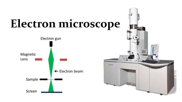
Electron Microscopy (EM)
Electron microscopy (EM) is a technique for obtaining high resolution images of biological and non-biological specimens.
Uses:
- It is used in biomedical research to investigate the detailed structure of tissues, cells, organelles and macromolecular complexes.
- The high resolution of EM images results from the use of electrons (which have very short wavelengths) as the source of illuminating radiation.
- Electron microscopy is used in conjunction with a variety of ancillary techniques (e.g thin sectioning, immuno-labeling, negative staining) to answer specific questions. EM images provide key information on the structural basis of cell function and of cell disease.
Types:
There are two main types of electron microscope –
Transmission EM (TEM)
The transmission electron microscope is used to view thin specimens (tissue sections, molecules, etc) through which electrons can pass generating a projection image. The TEM is analogous in many ways to the conventional (compound) light microscope.
TEM is used, among other things, to image the interior of cells (in thin sections), the structure of protein molecules (contrasted by metal shadowing), the organization of molecules in viruses and cytoskeletal filaments (prepared by the negative staining technique), and the arrangement of protein molecules in cell membranes (by freeze-fracture).
Scanning electron microscope (SEM)
The scanning electron microscope used a technique known as raster scanning to produce magnified images of the specimen. It directs a focused electron beam across the rectangular area of the specimen, which loses energy as it passes through. The energy is converted into other forms of energy, such as heat, light, secondary electrons, and backscattered electrons. This information can be translated to view the topography and composition of the original specimen.
![]()