Today our topic of discussion is ” Gross Anatomy of the Kidney “. The kidneys are a pair of bean-shaped organs that play a vital role in the urinary system. They are essential for filtering waste products, excess substances, and liquids from the bloodstream to form urine.
This article delves into the gross anatomy of the kidney, outlining its structural features, location, blood supply, and functional relevance. Understanding the gross anatomy of the kidneys is crucial for comprehending how the urinary system maintains homeostasis and provides insights into the diagnostics and treatment of renal diseases.
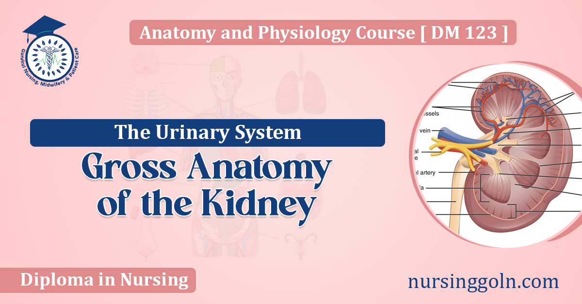
Gross Anatomy of the Kidney : The Urinary System
Location and External Anatomy
Position in the Body
- Retroperitoneal placement
- The vertebral level (T12 to L3)
- Relation to other organs (liver, intestines, spleen)
Size and Shape
- Average dimensions
- External landmarks (hilum, poles)
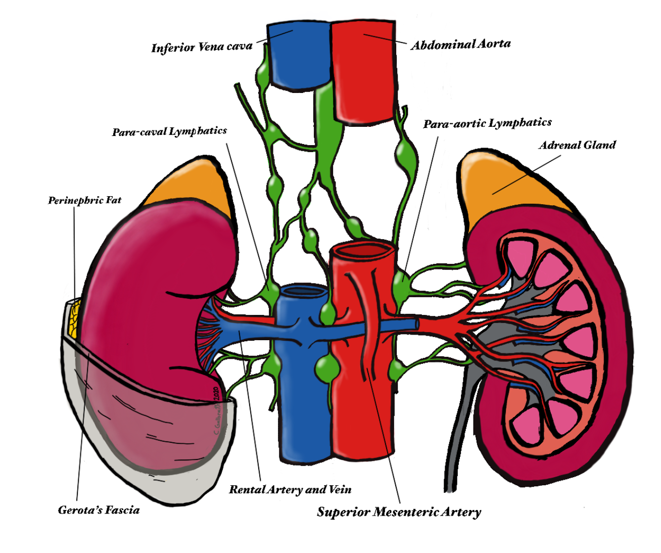
Protective Coverings
- Renal capsule
- Perirenal fat
- Renal fascia

Internal Anatomy
Renal Hilum
- Entry and exit point for structures (ureter, blood vessels, nerves)
- Structural arrangement
Renal Cortex and Medulla
- Texture and color differentiation
- Renal columns and pyramids
- Corticomedullary junction significance
Renal Papillae and Calyces
- Papillary ducts
- Minor and major calyces formation
- Flow into the renal pelvis
Renal Pelvis
- Funnel-shaped structure
- Transition to the ureter
Blood Supply and Lymphatics
Arterial Supply
- Renal artery branching
- Segmental arteries
- Interlobar, arcuate, and interlobular arteries
Venous Drainage
- Renal vein and its tributaries
- The route of blood back to the inferior vena cava
Lymphatic System
- Lymph vessels and nodes associated
- Role in homeostasis and immune response
Nervous Innervation
Sympathetic and Parasympathetic Fibers
- Source (renal plexus, celiac plexus)
- Regulatory function
Sensory Innervation
- Pain perception
- Reflex arcs
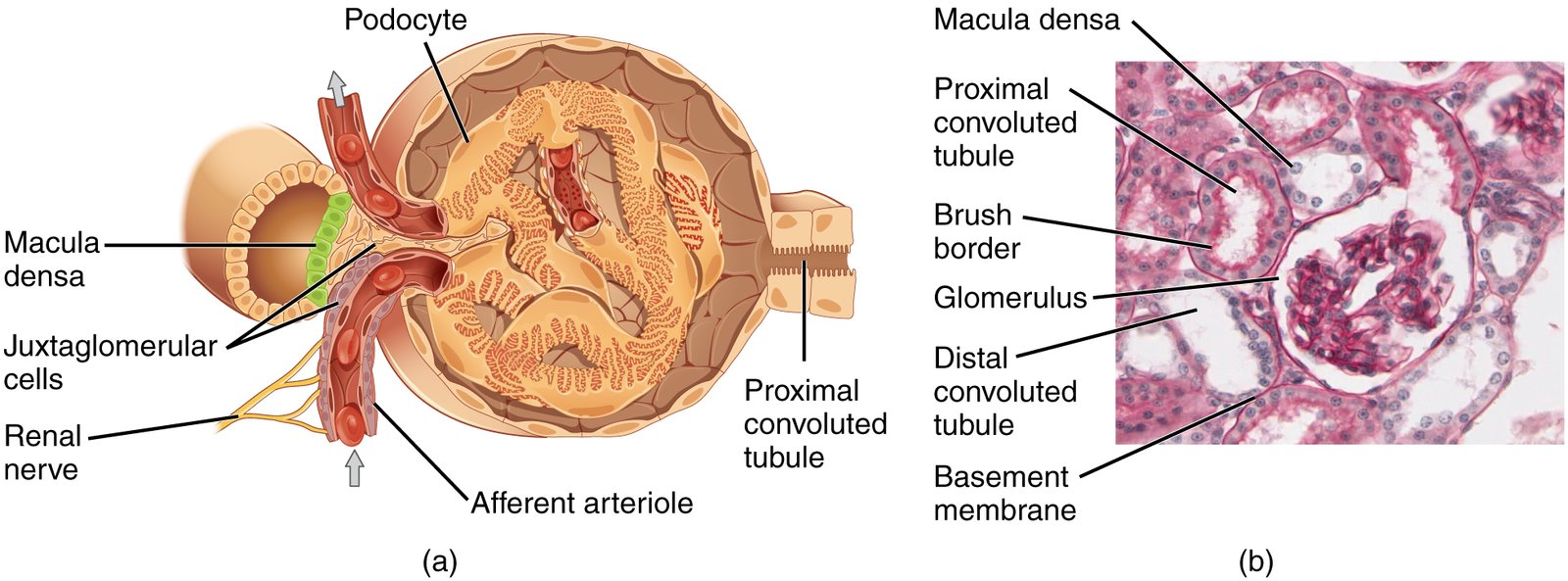
Microscopic Anatomy
Nephrons
- Structural and functional units
- Renal corpuscle (glomerulus and Bowman’s capsule)
- Renal tubule (proximal tubule, loop of Henle, distal tubule)
Collecting System
- Collecting tubules
- Collecting ducts
- Role in urine concentration
Physiological Relevance
Filtration, Reabsorption, Secretion
- Processes within the nephron
- Homeostatic regulation (fluid, electrolytes, acid-base balance)
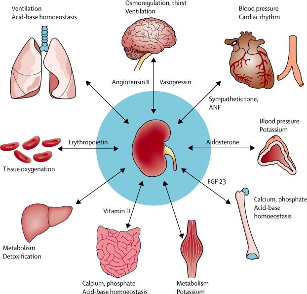
Hormonal Influence
- Renin-angiotensin-aldosterone system
- Antidiuretic hormone
Metabolic Functions
- Erythropoietin production
- Activation of vitamin D
Developmental Anatomy
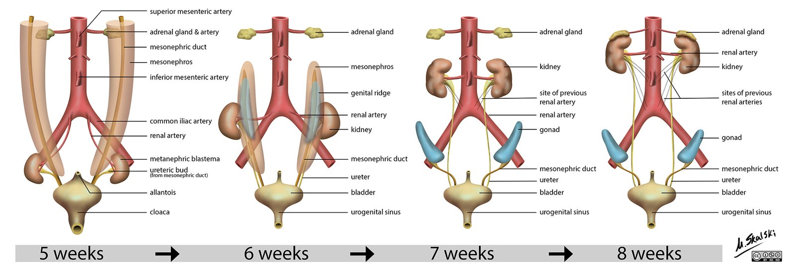
Embryological Origin
- Pronephros, mesonephros, and metanephros development stages
- Timing and clinical significance
Congenital Anomalies
- Polycystic kidney disease
- Horseshoe kidney
- Duplex collecting system
Clinical Correlations
Renal Pathologies
- Acute and chronic renal failure
- Glomerulonephritis
- Kidney stones
Diagnostic Procedures
- Ultrasonography
- CT/MRI scans
- Renal biopsy
Surgical Considerations
- Nephrectomy
- Transplantation
- Renal artery stenting
Conclusion
The kidneys’ gross anatomy is both intricate and elegant, mirroring their complex physiological tasks. A profound understanding of renal structure is indispensable for the medical community, as it underpins every aspect of renal function from basic physiology to clinical pathology.
The kidneys not only cleanse the blood of waste products and toxins but also maintain the delicate balance of fluids and electrolytes, making them critical to the survival of the organism. Knowledge of kidney anatomy is, therefore, a cornerstone of medical education, underpinning the diagnosis, management, and treatment of renal diseases.