Head Injury – An orthopedic nurse is a nurse who specializes in treating patients with bone, limb, or musculoskeletal disorders. Nonetheless, because orthopedics and trauma typically follow one another, head injuries and infected wounds are frequently treated by orthopedic nurses.
Ensuring that patients receive the proper pre-and post-operative care following surgery is the responsibility of an orthopedic nurse. They play a critical role in the effort to return patients to baseline before admission. Early detection of complications following surgery, including sepsis, compartment syndrome, and site infections, falls under the purview of orthopedic nurses.
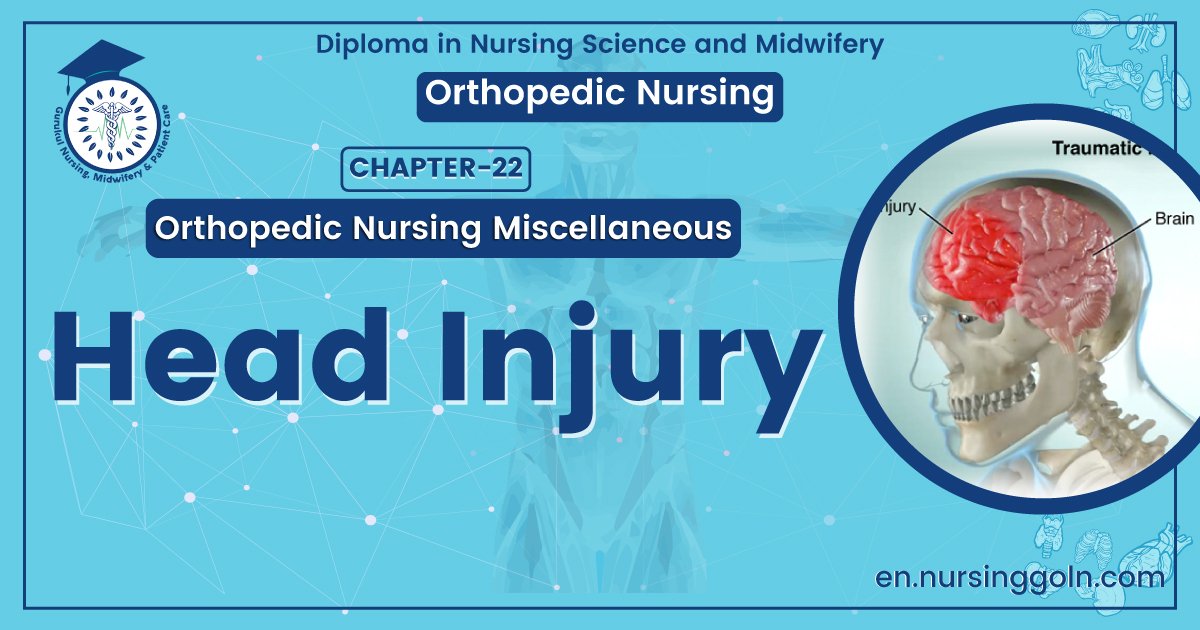
Head Injury
Traumatic brain injury:
Central nervous system (CNS) injury is the most common cause of death from injury. Two million people per year in the United States suffer traumatic brain injuries (TBIs), many as the result of motor vehicle crashes and falls. Approximately 50,000 deaths per year and 500,000 hospital admission are attributable to head injury.
Most of these victims are Between the ages of 16 and 30 years. The increasing use of seat belts and airbags has resulted in an estimated 20% to 25% reduction in these traffic fatalities. However, the incidence of penetrating injury to the brain and spinal cord is increasing. As awareness of the correct methods for brain injury management grows, guidelines for TBI have developed and been shown to improve outcome.

Classification of head injury:
TBIs are categorized as mild (80%), moderate (10%), or severe (10%), depending on the level of neurologic dysfunction at the time of initial evaluation. Determination of the Glasgow Coma Scale (GCS) score as early as possible and then serially is essential. Loss of consciousness (LOC) is an important indicator of TBI. Classification of TBI is based on the GCS.
A. Mild head injury:
1. GCS score of 13 to 15
2. Brief period of LOC
3. Prognosis is excellent
4. Mortality rate <1%
B. Moderate head injury:
1. GCS score of 9 to 12
2. Typically, confused and may have focal neurologic deficits; able to follow simple commands
3. Prognosis is good. 4. Mortality rate <5%
C. Severe head injury:
1. GCS of ≤8-generally, the accepted definition of coma
2. Unable to follow commands
3. Until recently, mortality >40%
4. Most survivors have significant disabilities
5. Airway control is essential
6. Elevated ICP is a common cause of death and neurologic disability
Signs and Symptoms of traumatic head injury:
The signs and symptoms of head traumas are:
i. persistent headaches,
ii. nausea,
iii. Loss of consciousness.
iv. Blurred vision,
v. Slurred speech,
vi. Memory problems are also high on the list

Manage a case of traumatic head injury:
A. Initial evaluation and treatment of head injury:
General:
1. Patients suspected of having suffered a head injury, particularly if confused or unresponsive, require emergency evaluation and treatment at a center with capabilities for immediate neurosurgical intervention. General objectives are:
- Rapid diagnosis and evacuation of intracranial mass lesions.
- Expedient treatment of extracranial injuries.
- Avoidance of secondary brain injury due to hypoxia and hypotension.
- Assess the history of other secondary insults such as hyperglycemia, hypothermia, and anemia may also exacerbate outcome during the hospital course.
2. Severe brain injury is associated with cerebral ischemia. Therefore, a principal therapeutic goal is to enhance cerebral perfusion and oxygenation and avoid further ischemic injury to the brain.

B. Initial management of the unresponsive patient with head injury:
Intubation with controlled ventilation (avoid routine hyperventilation) with assessment of GCS, pupillary response, and all four extremity movement to avoid pharmacologic paralysis. Venous access: Restore intravascular volume, blood pressure, and perfusion. Avoid hypotonic or dextrose-containing solutions. Immobilize the patient with rigid backboard and cervical spine (C-spine) collar. Pharmacologic paralysis and sedation, if agitated or combative Short-acting agents are recommended. Vecuronium bromide, cisatracurium, or succinylcholine Opioid sedation: fentanyl or morphine Avoid benzodiazepines Monitor blood pressure and O₂ saturation continuously. Check arterial blood gases (ABG), blood glucose, electrolytes, prothrombin time (PT), partial thromboplastin time (PTT), hematocrit, and platelet count. With active therapy, serum sodium levels and osmolality should be tracked.
C. Initiate medical management of the head injury: Proceed with rapid acquisition of a computed tomographic (CT) scan of the head and complete cervical spine (if time permits). Based on time, distance, and local capabilities, transfer may be necessary Rapid referral to a center capable of immediate neurosurgical intervention may be required. Do not delay transport to definitive care to obtain a CT scan of the head. Early diagnosis and evacuation of cranial mass lesions are critical. Repeated neurologic examination and assessment of GCS. Documentation of the GCS in patients who are intubated, or “tubed,”.
Hyperventilation causes cerebral vasoconstriction and can worsen cerebral ischemia. Routine hyperventilation should no longer be used. Hyperventilation is indicated only in the setting of abrupt neurologic deterioration with suspected herniation. C. Secondary management The avoidance of secondary brain injury is essential. Secondary brain injury is produced by hypoxia and hypotension.

A single episode of hypotension (systolic blood pressure <90 mmHg) in the adult will worsen prognosis and can increase mortality up to 50%. GCS obtained in the emergency department may be a more reliable assessment of the severity of brain The injury than the GCS obtained in the field. The GCS cannot be assessed by simple observation and requires stimulation of the patient. In cases of asymmetry in either eye opening or motor scores, the best score is used. A lateral cervical spine x-ray study usually can be obtained during secondary survey of the patient to detect gross injury or malalignment of the cervical spine
D. Indications for ICP monitoring. As a general approach, liberal use of ICP monitoring in patients with severe TBI (GCS ≤8) is recommended. An ICP monitor should also be considered in a patient with moderate head injury who is going to the OR for other injuries. ICP monitoring is indicated for: Severe closed head injury (GCS ≤8) and abnormal CT of head Definition of abnormal CT:
- Hematoma
- Contusion
- Edema
- Compressed basal cisterns
Severe closed head injury (GCS ≤8) and normal CT of head, particularly if two or more of the following exist:
- Age >40 years
- Unilateral or bilateral flexor or extensor posturing
- Systolic blood pressure <90 mmHg (rapid correction of hypotension is essential)
E. Intensive care management of patients with severe TBI (GCS ≤8). The goal is to prevent secondary brain injury by limiting focal cerebral ischemia, preventing cerebral hypoxia and maintaining adequate cerebral perfusion. This can be accomplished only by the continuous monitoring of several physiologic parameters and the judicious use of therapies to lower elevated ICP.
- Recommendations for physiologic monitoring of the patient with severe TBI:
- Arterial blood pressure. Noninvasive monitoring can be used, but an arterial catheter is preferred.
- Heart rate, electrocardiogram (ECG), temperature, and pulse oximetry.
- Central venous pressure or pulmonary artery catheter monitoring if the patient’s volume status is in question.
- ICP monitoring.
- Brain tissue O, (and if available, cerebral microdialysis).
- Fluid balance (intake and output).
- Arterial blood gases every 4 to 6 hours initially; electrolytes, glucose, and serum osmolality (if receiving mannitol) every 6 hours; hematocrit, PT, PTT, platelets every 12 hours.
- Jugular venous O₂ saturation or O, content by local protocol if hyperemia.
Read more: