Today our topic of discussion is ” Heart Anatomy “. The cardiovascular system, often visualized as an extensive network of vessels, is anchored by its central pump: the heart. This muscular organ’s intricate anatomy enables it to effectively circulate blood, oxygen, and nutrients throughout the body, ensuring optimal function of every tissue and organ. This article delves deep into the heart’s anatomy, providing a comprehensive understanding of its structure and significance.
Heart Anatomy – The Cardiovascular System: The Heart
1. Anatomical Position and External Features
The heart, roughly the size of a clenched fist, resides in the mediastinum, the central compartment of the thoracic cavity. It’s nestled between the lungs, positioned slightly left of the midline, with its apex pointing downward and to the left.
2. Heart Wall: The Three Layers
a. Epicardium (Visceral Pericardium): The heart’s outermost layer is a thin sheet of serous membrane, which also extends to form the parietal layer of the pericardium. b. Myocardium: This thick, muscular middle layer comprises cardiac muscle tissue, responsible for the heart’s contractions. c. Endocardium: The innermost layer, a thin endothelium, lines the heart chambers and continues into the blood vessels.
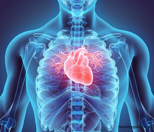
3. Chambers of the Heart
The heart is divided into four chambers:
a. Atria (Upper Chambers):
- Right Atrium: Receives deoxygenated blood from the body via the superior and inferior vena cava.
- Left Atrium: Gathers oxygen-rich blood from the lungs via the pulmonary veins.
b. Ventricles (Lower Chambers):
- Right Ventricle: Pumps the deoxygenated blood into the lungs through the pulmonary artery.
- Left Ventricle: The heart’s most muscular chamber, it drives oxygen-rich blood into the systemic circulation through the aorta.
4. Heart Valves: The Flow Regulators
Ensuring unidirectional blood flow, the heart houses four valves:
a. Atrioventricular (AV) Valves: Positioned between the atria and ventricles.
- Tricuspid Valve: Located between the right atrium and right ventricle.
- Bicuspid or Mitral Valve: Sits between the left atrium and left ventricle.
b. Semilunar Valves: Found between the ventricles and the large arteries.
- Pulmonary Valve: Connects the right ventricle to the pulmonary artery.
- Aortic Valve: Links the left ventricle to the aorta.
5. The Electrical Conduction System
The heart’s rhythmic contractions stem from its unique electrical conduction system:
a. Sinoatrial (SA) Node: Often termed the heart’s natural pacemaker, it initiates electrical impulses, which then spread throughout the atria, making them contract.
b. Atrioventricular (AV) Node: Located in the right atrium, it briefly delays the impulse, allowing the ventricles to fill with blood.
c. AV Bundle (Bundle of His) and Bundle Branches: Transmit the impulse from the AV node to the ventricles.
d. Purkinje Fibers: These fibers spread the impulse to the myocardium of the ventricles, causing them to contract.
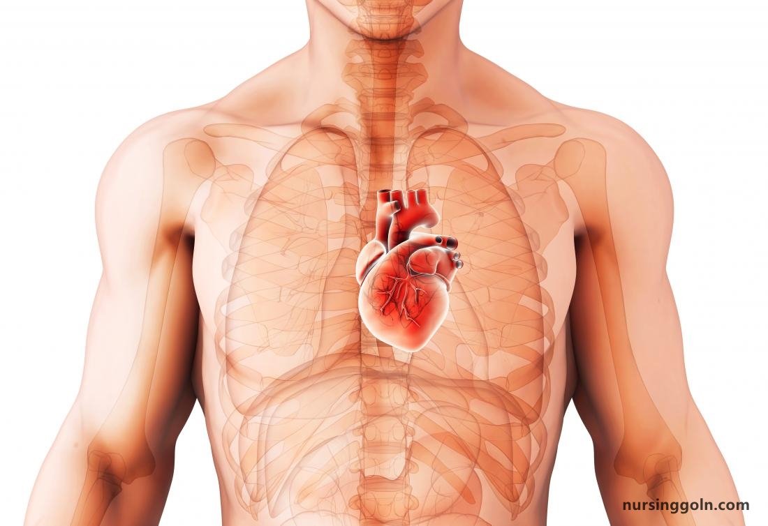
6. Blood Supply to the Heart: The Coronary Circulation
The heart’s muscle, despite being in constant contact with blood, receives its nourishment from the coronary arteries:
a. Right and Left Coronary Arteries: Originate from the base of the aorta, branching out to supply various heart regions. b. Cardiac Veins: Collect the deoxygenated blood and channel it into the coronary sinus, which empties into the right atrium.
7. The Protective Sac: The Pericardium
Surrounding the heart is a double-layered sac called the pericardium. It offers protection and reduces friction as the heart beats:
a. Fibrous Pericardium: The tough, outer layer. b. Serous Pericardium: The inner layer, further divided into the parietal and visceral layers, with a cavity (filled with pericardial fluid) in between for lubrication.
8. The Heart’s Lifelong Journey
From its first beat in the fetus to its last, the heart undergoes various changes:
a. Fetal Circulation: Structures like the foramen ovale and ductus arteriosus allow blood to bypass the lungs, which aren’t functional before birth. b. Age-related Changes: Over time, the heart can experience decreased efficiency, thickening of its walls, or reduced elasticity of the valves.
Conclusion
The heart’s intricate anatomy showcases nature’s brilliance, ensuring optimal efficiency in oxygen and nutrient delivery. Each chamber, valve, and vessel plays a pivotal role in sustaining life. Recognizing the significance of heart health, it becomes evident why proactive measures to care for this organ are not just recommended but essential.