Hypercapnia, hypoxia Cyanosis –The course is designed for the basic understanding of anatomical structures and physiological functions of human body, musculoskeletal system, digestive system, respiratory system; cardiovascular system; urinary system, endocrine system, reproductive system, nervous system, hematologic system, sensory organs, integumentary system, and immune system.The aim of the course is to acquire knowledge and skills regarding anatomy and physiology.
Hypercapnia, hypoxia Cyanosis
Hypercapnia
Definition: Hypercapnia means excessive CO2 concentration in blood or body fluid.
Causes:
- Hypovetilation (common cause): Due to poisoning of drugs e.g. morphine, barbiturates, brain stroke/hemorrhage
- Obstruction in the respiratory passage.
- Circulatory deficiency.
Clinical feature:
- Confusion
- Diminished sensory acuity
- Coma with respiratory depression and death.
Treatment:
The patient should be treated with high concentration (> 35%) oxygen by Oro-nasal mask
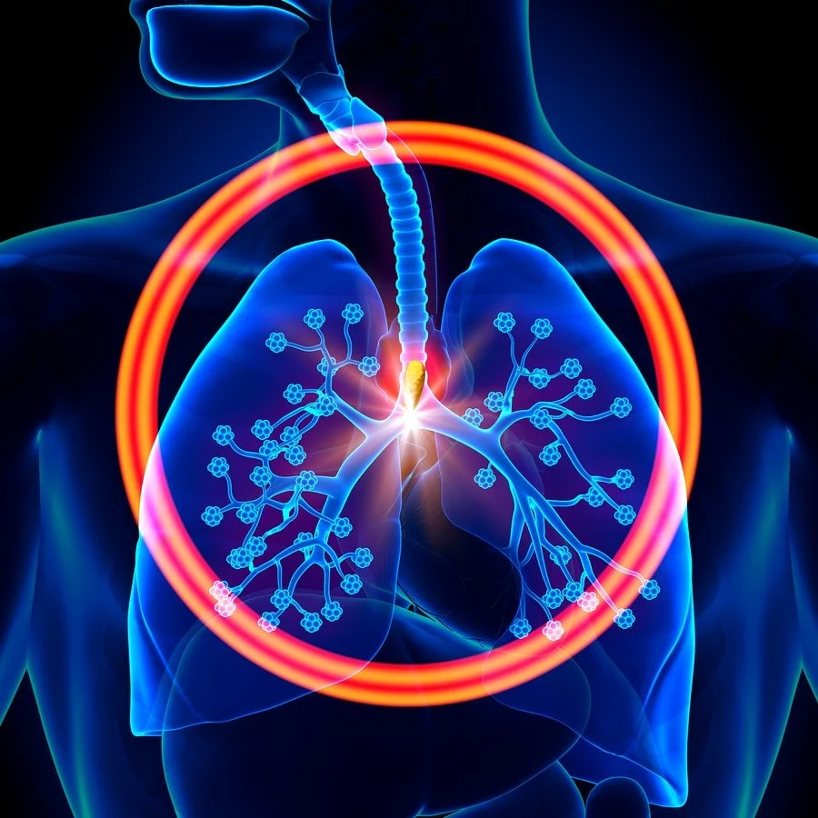
Hypoxia
Definition:
Hypoxia is oxygen (O2) deficiency at the tissue level.
Types with causes:
- Нурокіc hypoxia (anoxic anoxia): In which the PO2 of the arterial blood is reduced.
- Anaemic hypoxia: In which the arterial PO2 in normal but the amount of haemoglobin available to carry O2 is reduced
- Stagnant or ischemic hypoxia: In which the blood flow to a tissue is so low that adequate O2 is not delivered to it despite a normal PO2 and Hb, concentration.
- Histotoxic hypoxia: In which the amount of O2 delivered to tissue is adequate but the tissue cannot utilize the 02 due to presence of toxic substances.
(Ref-Ganong 23/617p)
Asphyxia
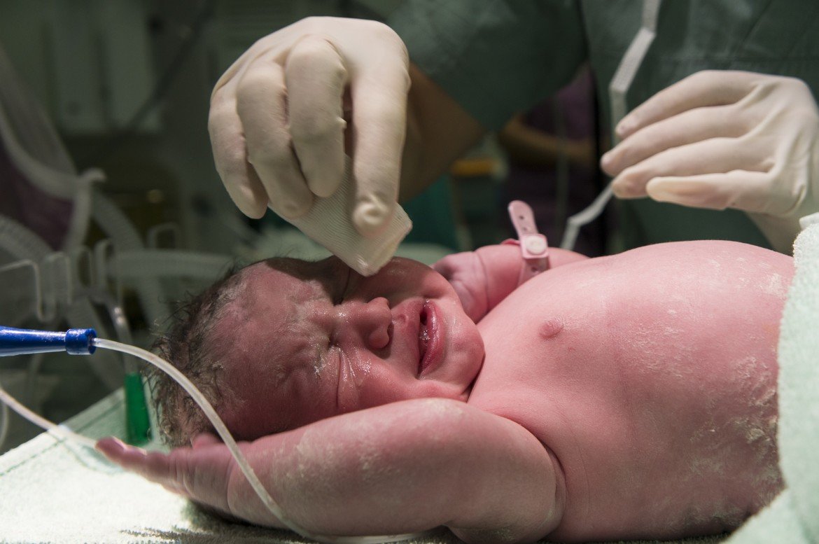
It is the condition caused by insufficient intake of oxygen.
Causes:
- Occlusion of the airways.
- Acute hypercapnea
Effects:
- There is marked stimulation of respiration with violent respiratory effort
- Blood pressure and heart rate rise sharply
- Catecholamine secretion is increased
- Blood pH falls
Management:
Artificial respiration. If artificial respiration is not started immediately, cardiac arrest occur within 4-5 minutes
Oxygen (O) therapy
Administration of O, rich gas mixture to increase in the amount of dissolved O, in the arterial blood.
O, therapy has a great value in hypoxic hypoxia except in that case where hypoxic hypoxia is due to shunting of un-oxygenated venous blood bypassing the lungs. It has also value in stagnant, anemic and histotoxic hypoxia but less amount
Indications of O2 Therapy:
- Shock.
- Hypoxia
- CO poisoning
- Pneumonia.
- Respiratory distress
- Pulmonary edema
- Obstructive lung diseases
- Myocardial infarction etc
The effects of O_{2} toxicity:
Administration of O_{2} may stop respiration and even causes death in a patient with severe respiratory failure because, high concentration of O, (100% O_{2} ) can cause tissue damage by producing free radicals (O 2 \& H_{2}*O_{2} )
The toxicities that develop after giving O_{2} 80-100% O_{2} for 8 hours or more) are-
- Respiratory tract: Irritation, nasal congestion, sore throat, coughing etc.
- Lungs: Pulmonary edema, atelactasis, hemorrhage etc.
- Brain: Muscle twitching, dizziness, convulsion, coma and which may lead to death.
(Ref: Ganong 23 622p.)
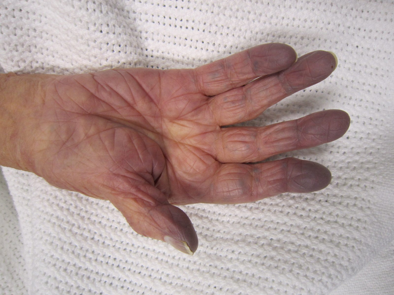
Cyanosis
Definition:
Cyanosis may be defined as bluish coloration of the skin & mucous membrane due to excess amount of reduced hemoglobin (Deoxygenated Hb) in the blood (more than 5gm / d * l )
Cause:
- Hypoxic hypoxia.
- Stagnant hypoxia.
Site of observation:
- Nail bed
- Mucous membrane.
- Ear lobules
- Lips & finger,
- Tip of the tongue & nose.
Types of cyanosis:
- Central cyanosis.
- Peripheral cyanosis
Treatment:
Removing hypoxia by O_{2} therapy, In case of obstructive lung disease: O_{2} & C*O_{2} therapy
(Ref-Guyton and Hall 12 ^ (ck) * 521p .)
Chronic mountain sickness
If a person remains at high altitude for a long time, he develops chronic mountain sickness
Feature of chronic mountain sickness:
- ↑red cell mass and hematocrit.
- Of pulmonary arterial pressure.
- Enlargement of right heart.
- peripheral arterial pressure
- Congestive cardiac failure.

Pulse oximetry
Noninvasive method of estimating the percentage of oxygen saturation in the arterial blood using an oximeter with a specialized probe attached to the skin at a site of arterial pulsation, commonly the finger. used to monitor hypoxemia
or,
A pulse oximeter is commonly used in hospitals for noninvasively measuring oxyhemoglobin saturation. This device typically clips on the finger or pinna and gives readings of oxygen saturation and pulse rate within a short time, making it useful in many clinical contexts such as in emergency medicine and during anesthesia. The normal value for arterial blood is about 97%
Read more: