Macroscopic Structures of Long Bone – The course is designed for the basic understanding of anatomical structures and physiological functions of human body, musculoskeletal system, digestive system, respiratory system; cardiovascular system; urinary system, endocrine system, reproductive system, nervous system, hematologic system, sensory organs, integumentary system, and immune system. The aim of the course is to acquire knowledge and skills regarding anatomy and physiology.
Macroscopic Structures of Long Bone
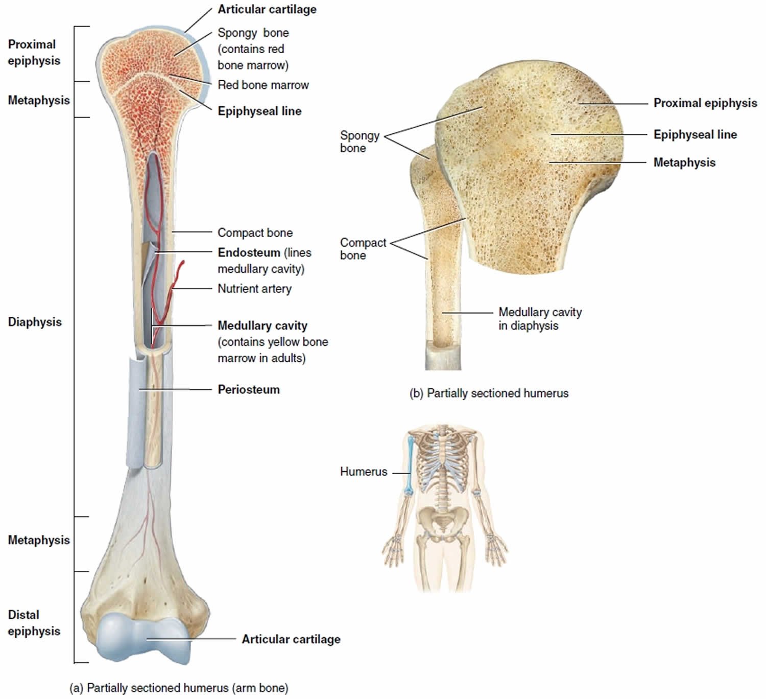
Macroscopic Structures of long Bone (Humerus)
The structure of a bone may be analyzed by considering the parts of a long bone, for instance, the humerus (the arm bone). A typical long bone consists of the following seven parts:
- The diaphysis
- The epiphyses
- The metaphyses
- The articular cartilage
- The periosteum
- The medullary
- The endosteum
Shortly description of Macroscopic Structures of long Bone
- The diaphysis (growing between) is the bone’s shaft or body-the long, cylindrical, main portion of the bone.
- The epiphyses (growing over; singular is epiphysis) are the distal and proximal ends of the bone.
- The metaphyses(meta- between; singular is metaphysis) are the regions in a mature bone where the diaphysis joins the epiphyses. In a growing bone, each metaphysis contains an epiphyseal (growth) plate, a layer of hyaline cartilage that allows the diaphysis of the bone to grow in length. When bone growth in length stops, the cartilage in the epiphyseal plate is replaced by bone and the resulting bony structure is known as the epiphyseal line.
- The articular cartilage is a thin layer of hyaline cartilage covering the part of the epiphysis where the bone forms an articulation (joint) with another bone. Articular cartilage reduces friction and absorbs shock at freely movable. joints. Because articular cartilage lacks a perichondrium, repair of damage is limited.
- The periosteum (peri = around) is a tough sheath of dense irregular connective tissue and its associated blood vessels that surrounds the bone surface wherever it is not covered by articular cartilage. The periosteum contains bone-forming cells that enable bone to grow in diameter or thickness, but not in length. It also protects the bone, assists in fracture repair, helps nourish bone tissue, and serves as an attachment point for ligaments and tendons.
- The medullary cavity (medulla marrow, pith) or marrow cavity is a hollow, cylindrical space within the diaphysis that contains fatty yellow bone marrow in adults.
- The endosteum (endo = within) is a thin membrane that lines the medullary cavity. It contains a single layer of bone-forming cells.
(Ref:- Guyton and Hall, Textbook of Medical Physiology. 12th ed + J. TORTORA, The essentials of anatomy and physiology, 8th edition, P-119,120)
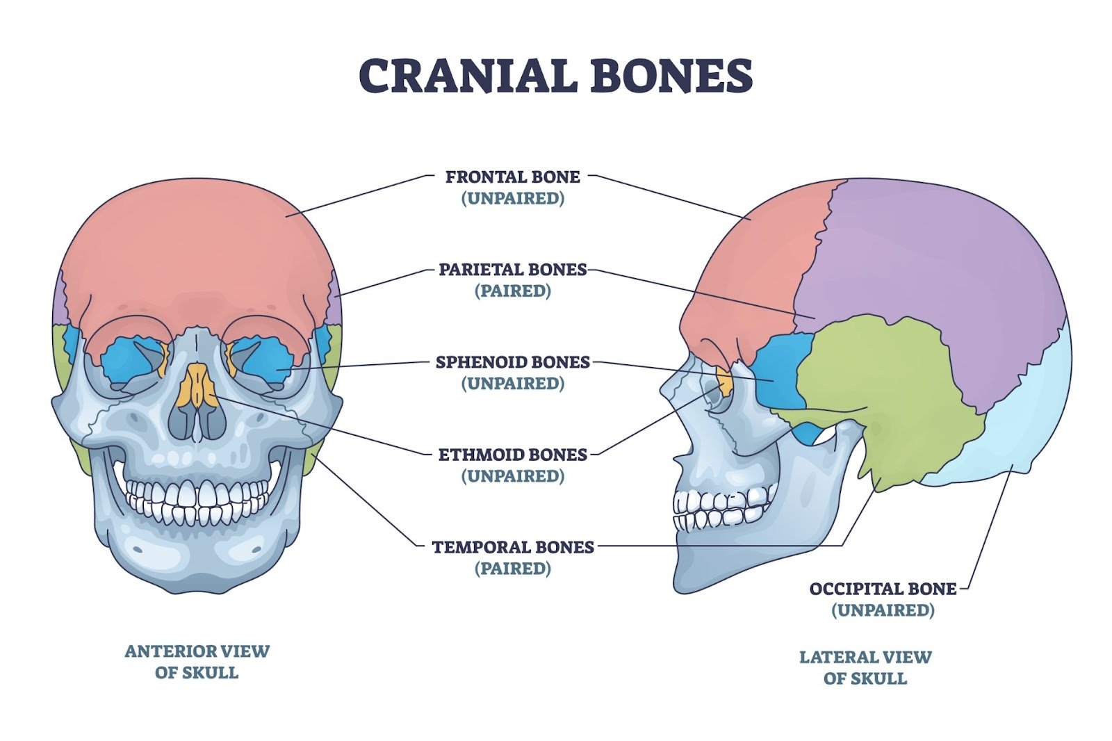
The skull, which contains 22 bones. It includes two sets of bones: cranial bones and facial bones.
The eight cranial bonesform the cranial cavity that encloses and protects the brain. They are:-
Frontal bone — —1
Parietal bones —–2
Temporal bones — 2
Occipital bone —-1
Sphenoid bone— 1
Ethmoid bone —- 1
_____________________________
Total = 8 bones
Fourteen facial bones form the face. They are:-
Nasal bones————2
Maxillae—————- 2
Zygomaticbones——- 2
the mandible———–1
Lacrimal bones——– 2
Palatine bones——— 2
Inferior nasal conchae- 2
Vomer——————-1
__________________________________
Total = 14 bones