Medical imaging represents one of the great marvels of modern medicine. It allows clinicians to peer inside the human body without the need for invasive procedures, providing invaluable insights into the structure and function of our internal organs, bones, and tissues. This article delves into the world of medical imaging, offering an introduction to various techniques, their applications, and how they revolutionize our understanding of the human body.
The Evolution of Medical Imaging
The history of medical imaging dates back to the late 19th century when Wilhelm Conrad Röntgen discovered X-rays in 1895. This marked a major turning point, enabling doctors for the first time to view the inside of the body without surgery. Over the subsequent century, numerous imaging techniques emerged, each offering unique insights into human anatomy and physiology.
Techniques in Medical Imaging
Medical imaging encompasses a range of techniques, each suited to different clinical needs. Some of the most widely used include:
1. X-ray Imaging (Radiography)
This is the oldest and most common form of medical imaging. When X-ray beams pass through the body, dense structures like bones absorb them, while softer tissues allow them to pass through. The resulting images, or “radiographs,” show these dense structures in detail, making them particularly useful for diagnosing fractures, infections, and tumors in bones.
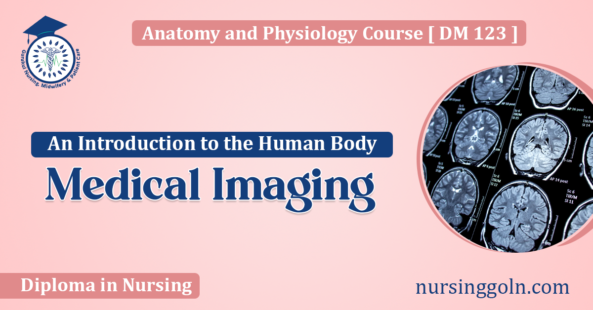
2. Magnetic Resonance Imaging (MRI)
MRI utilizes strong magnets and radio waves to create detailed images of soft tissues within the body. Unlike X-rays, MRI doesn’t use ionizing radiation. This imaging method is especially useful for visualizing the brain, muscles, and joints, as well as tumors in soft tissues.
3. Computed Tomography (CT) Scan
A CT scan combines multiple X-ray images taken from different angles to produce cross-sectional images of the body. These “slices” can be viewed individually or combined to create a 3D image. CT scans offer detailed views of bones, blood vessels, and soft tissues, making them essential for detecting tumors, hemorrhages, and other abnormalities.
4. Ultrasound Imaging
Also known as sonography, ultrasound uses high-frequency sound waves to produce images of structures within the body. A device called a transducer emits these sound waves, which bounce back after hitting internal structures. The echoes are then converted into images. Ultrasound is commonly used during pregnancy, as well as for visualizing the heart, blood vessels, and organs in the abdomen.
5. Positron Emission Tomography (PET) Scan
PET scans measure metabolic or chemical activity in tissues. Patients are given a small amount of a radioactive substance, which tissues absorb differently. When tissues break down this substance, they emit particles called positrons, detected by the PET scanner. This technique is often used to detect cancer, assess brain disorders, or study the heart.
The Role of Medical Imaging in Modern Medicine
Medical imaging is indispensable in contemporary healthcare. Its applications are vast, encompassing:
- Diagnostics: Imaging helps clinicians identify diseases, infections, and abnormalities, offering a definitive diagnosis.
- Treatment Planning: Imaging guides clinicians in developing personalized treatment plans, whether it’s determining a tumor’s size or planning surgical procedures.
- Monitoring: Medical imaging tracks the progression of diseases or the efficacy of treatments, providing real-time feedback to healthcare providers.
- Research: Medical researchers utilize imaging to study diseases, test new treatments, and delve deeper into the intricacies of human physiology.
Safety in Medical Imaging
While medical imaging has revolutionized healthcare, it’s essential to address concerns about safety, especially with techniques that use ionizing radiation, like X-rays and CT scans.
Radiation doses are usually kept as low as possible, adhering to the “As Low As Reasonably Achievable” (ALARA) principle. It’s crucial to ensure that the benefits of imaging outweigh potential risks, and patients should always be informed about these risks.
MRI, being radiation-free, poses different challenges. The strong magnetic fields can interact with metallic implants or devices within the body, necessitating careful screening before the procedure.
The Future of Medical Imaging
Advances in technology and computational methods are pushing the boundaries of medical imaging. Artificial intelligence (AI) is showing immense potential in enhancing image analysis, reducing errors, and speeding up diagnosis.
Additionally, hybrid imaging techniques, combining the strengths of two or more imaging methods, are emerging. For instance, combining PET with MRI (PET-MRI) offers metabolic insights from PET alongside the detailed anatomical images of MRI.
Furthermore, innovations like portable ultrasound devices and wearable imaging sensors could make imaging more accessible, especially in resource-limited settings.
Conclusion
Medical imaging provides a non-invasive window into the human body, transforming our ability to diagnose, treat, and study diseases. As we continue to innovate and refine these techniques, we move closer to a future where illnesses can be detected even before symptoms manifest, ensuring timely and effective interventions.
The human body is a complex, intricate system, and medical imaging offers a way to understand it better, one image at a time. As the world of medicine evolves, imaging will undoubtedly remain at its forefront, guiding clinicians and patients alike towards healthier futures.