Multiple pregnancy – This course is designed to understand the care of pregnant women and newborn: antenatal, intra-natal and postnatal; breast feeding, family planning, newborn care and ethical issues, The aim of the course is to acquire knowledge and develop competencies regarding midwifery, complicated labour and newborn care including family planning.
Multiple pregnancy
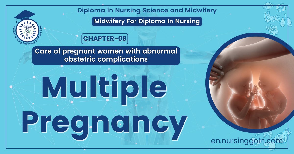
When more than one fetus simultaneously develops in the uterus, it is called multiple pregnancy.

Varieties of multiple pregnancy:
1. Twin (Commonest)
a. Monozygotic/uniovular/monovular/identical.
b. Dizygotic/binovular/fraternal
2. Quadruplets (four fetus).
3. Triplets (three fetus).
4. Quintuplets (five fetus)
5. Sextuple (six fetus)
Twin pregnancy:
Simultaneous development of two fetuses in the uterus is called twin pregnancy.
Varieties of twin pregnancy:
- Dizygotic twins (commonest and it is 2/7 of twins). It is resulting from the fertilization two ova.
- Monozygotic twins (1/3of twins).
Rarely:
- Diametric-diachronic twins.
- Diamnoitic monchorinic twins.
- Monoamniotic monochorionic twins.
- Conjoined twins.
Pathogenesis of twins (Dizygotic twins):
It is resulting from fertilization of two ova by two sperms during a single ovarian cycle.
Pathogenesis of twins (Monozygotic twins)
The twining may occur rat a different periods after fertilization.
Criteria of twins:
Dizygotic twins:
➤ There are two placentae
➤ Each fetus is surrounded by a separate amnion and chorion
➤ The intervening membranes consists of 4 layers (amnion and chorion and amnion)
Monozygotic twins:
➤ The placenta is single
➤ Each fetus is surrounded by separate amniotic sac with the chronic layer
➤ The intervening membranes consists of two layers of amnion only,
Factors responsible in multiple features:
➤ Idiopathic
➤ Iatrogenic: When ovulation inducing drugs are used
➤ Race: Highest among Negroes
➤ Hereditary
➤ Advancing age of the mother: Maximum in between 30-35 years
➤ Drugs: Ovulation inducing drugs
➤ Influence on parity

Conjoined twins:
When two fetus is connected to each other with some part of body is called conjoined twins.
Types: 4 types-
1. Thoracopagus (commonest)
2. Pyopagus (posterior fusion)
3. Craniopagus (cephalic)
4. Ischiopagus(caudal)
4 complications of conjoined of twins:
1. Over distension
2. Prolonged labour
3. Obstructed labour
4. Postpartum haemorrhage
The management of multiple pregnancy
Assessment:
➤ History of ovulation inducting drugs specially gonadotrophin, for infertility or use of ART.
➤ Family history of twining (more often present in the maternal side)
Symptoms:
Minor ailment of normal pregnancy is often exaggerated. Some of the symptoms are related to the undue enlargement of the uterus.
➤ Increased nausea and vomiting in early months.
➤ Cardio-respiratory embarrassment which is evident in the later months such as palpitation or shortness of breath.
➤ Tendency of swelling of the legs, varicose veins and hemorrhoids is greater.
➤ Unusual rate of abdominal enlargement and excessive fetal movements may be noticed by an experienced parous mother.
General examination:
➤ Prevalence of anaemia is more than in single pregnancy.
➤ Unusual weight gain, not explained by preeclampsia or obesity, is an important feature.
➤ Evidence of preeclampsia (25%) is a common association.
Abdominal examination:
Inspection:
➤ The elongated shape of a normal pregnant uterus is changed to a more “barrel shape” and the abdomen is unduly enlarged.
➤ The height of the uterus is more than the period of amenorrhoea.
➤ The girth of the abdomen at the level of umbilicus is more than the normal average at term (100 cm).
✓ Fetal bulk seems disproportionately large in relation to the size of the fetal head.
✓ Palpation of too many fetal parts.
➤ Finding of two fetal heads or three fetal poles make the clinical diagnosis almost certain..
Auscultation:
Simultaneous hearing of two district fetal heart sounds located at separate spots with a silent area in between by two observers, gives a certain clue in the diagnosis of twins, provided the difference in heart rate is at least 10 beat per minute.
Internal examination:
In some cases, one head is felt deep in the pelvic, white the other one i located by abdominal examination.
Investigation:
Ultrasonography to detect –
➤ 2 gestational sacs (at 10th weeks)
➤ 2 fetal heads at 14th weeks.
In multifetal pregnancy it is one to obtain the following information;
➤ Confirmation of diagnosis as early as 10th weeks of pregnancy.
➤ Viability of fetuses.
➤ Chorionicity (lambda or twin peak sign).
➤ Pregnancy dating.
➤ Fetal anomalies.
➤ Fetal growth monitoring (at every3-4 weeks interval) for IUGR.
➤ Presentation and lie of the fetuses.
➤ Twin transfusion (Doppler studies).
➤ Placental localization.
➤ Amniotic fluid volume.
Management during labour (Place of delivery: Hospital)
A. First stage:
Usual conduction of the first stage as outlined for a singleton fetus is to be followed with additional precautions:
a. A skilled obstetrical should be present.
b. The patient should be in bed to prevent early rupture of the membranes.
c. Use of analgesic drugs is to be limited as the babies are small and rapid delivery may occur.
d. Careful fetal monitoring is to be done with available gadgets.
e. Internal examination should be done soon after the rupture of the membranes to exclude cord prolapse
f. An intravenous line with ringer’s solution should be set up for any urgent intravenous therapy, if required.
g. One unit of compatible and cross matched should be made readily available.
h. Neonatologist should be present at the time of delivery.
B. 2nd stage:
The delivery should be conduct in the same guidelines as mentioned in normal labour:
a. Liberal episiotomy under local infiltration with 1% lignocaine.
b. Forceps delivery, if needed.
c. Do not give intravenous ergometrine with the delivery of the anterior shoulder of the first baby.
d. Clamp the cord at two places and cut in between.
e. At least 8-10 cm of cord is left behind for administration of any drug or transfusion, if required
f. The baby is handed over to the nurse after labeling it as number 1.

Delivery of the 2nd baby:
Principles:
Delivery of the 2nd baby without must be done without undue prolongation.
C. 3rd stage:
a. 2 ampoule oxytocin i/v/or i/m immediately just after delivery of the baby.
b. Controlled cord traction.
c. Fundal massage
d. The patient is to be watched carefully for about 2 hours after delivery.
Principles of delivery of 2nd baby:
Must be done without undue prolongation.
Indications of cesarean section for the 2nd baby:
- Larger second twin with noncephalic presentation.
- Prompt closure of the cervix after the delivery of the first baby
Complications of multiple pregnancy:
Maternal
During pregnancy:
➤ Nausea and vomiting.
➤ Anemia
➤ Pre-ecalmpsia
➤ Hydramnios
➤ Antepartum haemorrhage.
➤ Malpresentation
➤ Preterm labour
During labour:
➤ Early rupture of the membranes and cord prolapsed.
➤ Prolonged labour.
➤ Increased operative interference.
➤ Postpartum haemorrahage.
➤ Subinvolution
➤ Lactation failure
Fetal complication:
➤ Miscarriage
➤ Prematurely
➤ Intrauterine death.
➤ Fetal anomalies.
➤ Asphyxia and stillbirth.
➤ Pre-eclampsia
➤ Malpresentation
➤ Placental abruption.

Differential diagnosis of multiple pregnancy:
a) Hydramnios
b) Big baby
c) Fiborid or ovarian tumour with pregnancy.
d) Ascites with pregnancy.
Twin transfusion syndrome (TTS):
It is a clinic pathological state, exclusively met with in monozygotic twins where one twin appears to bleed into the other through some kind of placenta vascular anstomosis.
Clinical manifestations:
Occurs when there is haemodynamic imbalance due to unidirectional deep arteriovenous anastomose-
a) As a result the receptor twin becomes larger with hydramnios, polycythemic, hypertensive and hypervolaemic.
b) At the expensive of the donor twin which becomes smaller with oligohydramnios anemic, hypotensive and hypovolaemic.
c) The donor twin may appear “stuck” due to severe oligohydramnios.
d) Difference of haemoglobin concentration between the two, usually exceeds 5mg% and estimated fetal weight discrepancy is 25% or more.
e) The smaller twin generally have got better outcome
f) Perinatal mortality of TTS is about 70%
Diagnosis:
➤ By USG with droppler blood flow study in the placental vascular bed
Treatment:
a) Repeated amniocentesis: To control polyhydramnios in the receptor twin
b) Laser photocoagulation: To interrupt the anaestomotic vessels on the chorionic plate can give some success.
