
Pace maker of heart -The course is designed for the basic understanding of anatomical structures and physiological functions of human body, musculoskeletal system, digestive system, respiratory system; cardiovascular system; urinary system, endocrine system, reproductive system, nervous system, hematologic system, sensory organs, integumentary system, and immune system.The aim of the course is to acquire knowledge and skills regarding anatomy and physiology.
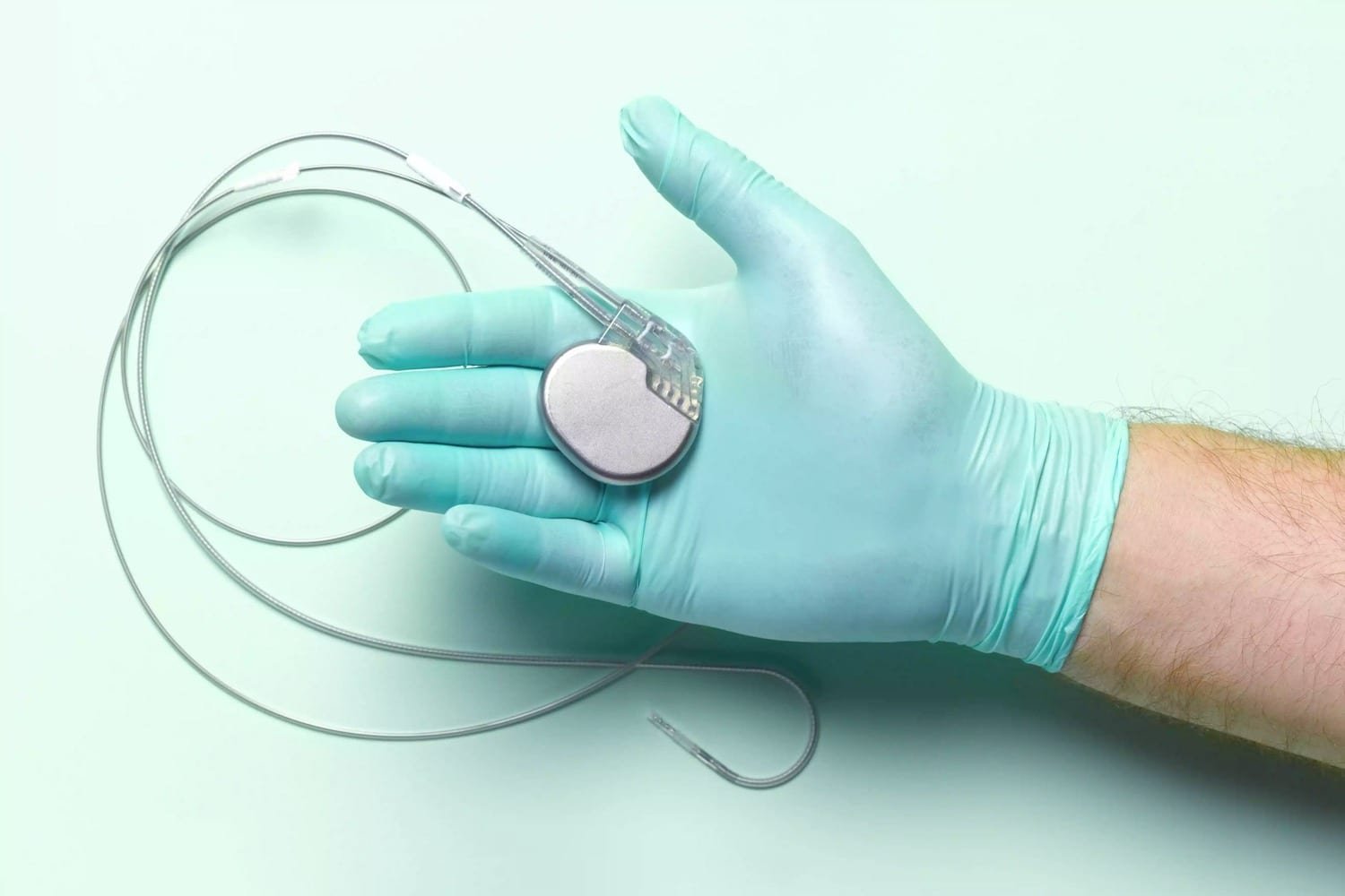
Pace maker of heart
Pacemaker:
A specialized cell or group of cells that automatically generates impulse that may spread to other pans at the heart is called pace maker
Pacemaker tissues of the heart:
- SA node.
- AV node
- Purkinje system.
SA node is called pace maker because:
- SA node generates cardiac impulse at first.
- It maintains normal cardiac rhythm.
- The rate and rhythm originated by SA node is higher than that of any other part of the heart
Importance:It maintains normal cardiac rhythmicity
[N.B: Pacemaker tissues have 3 types of cells-
- P-cells found in SA node & AV node
- Purkinje cells found in purkinje system
- Transitional cells.
Heart rate
Heart rate: The number of heart beats in a minute is called heart rate.
Normal heart rate in different age:
In ferus: 140-150/min.
In newborn : 130-140/min.
In children: 80-120/min
In adult :60-100/min
In old age :75-80/min
Tachycardia:
When the heart rate is above the normal physiological limit. Usually above 100 beats/minute.
Bradycardia:
When the heart rate is below the normal physiological limit.
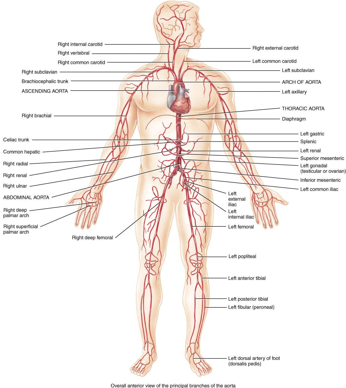
Common sites for examination of arterial pulse:
| Pulse | Common sites for examination. |
| Radial pulse | At the hand, just lateral to the tendon of the flexor carpi radialis muscle |
| Brachial pulse | At the elbow, just medial to the tendon of the biceps muscle. |
| Carotid pulse | At the neck, between the larynx & anterior border of the sternocleidomastoid muscle |
| The arteria dorsalis pedis | At the foot, just lateral to the tendon of the extensor hallucis longus. |
| The posterior tibial artery | Just behind the medial malleolus, midway between tendoAchllis. |
| The popliteal artery | Against the posterior aspect of the tibial condyle |
| The femoral artery | At the groin just below the inguinal ligament midway between the anterior superior iliac spine & the pubis symphysis. |
Cardiace output
The volume of blood ejected per minute from the left ventricle into the aorta is called the cardiac output (CO). (Note that the same amount of blood is also ejected from the right ventricle into the pulmonary trunk) Cardiac output is determined by
(1) the stroke volume (SV), the amount of blood ejected by the left ventricle during each beat (contraction), and
(2) heart rate (HR), the number of heartbeats per minute.
In a resting adult, stroke volume averages 70 mL, and heart rate is about 75 beats per minute. Thus the average cardiac output in a resting adult is Cardiac output = stroke volume X heart rate = 70 ml./beat X 75 beats/min 5250 ml/min or 5.25 liters/min Factors that increase stroke volume or heart rate, such as exercise, increase cardiac output.