Post-Partum Hemorrhage (PPH) – This course is designed to understand the care of pregnant women and newborn: antenatal, intra-natal and postnatal; breast feeding, family planning, newborn care and ethical issues, The aim of the course is to acquire knowledge and develop competencies regarding midwifery, complicated labour and newborn care including family planning.
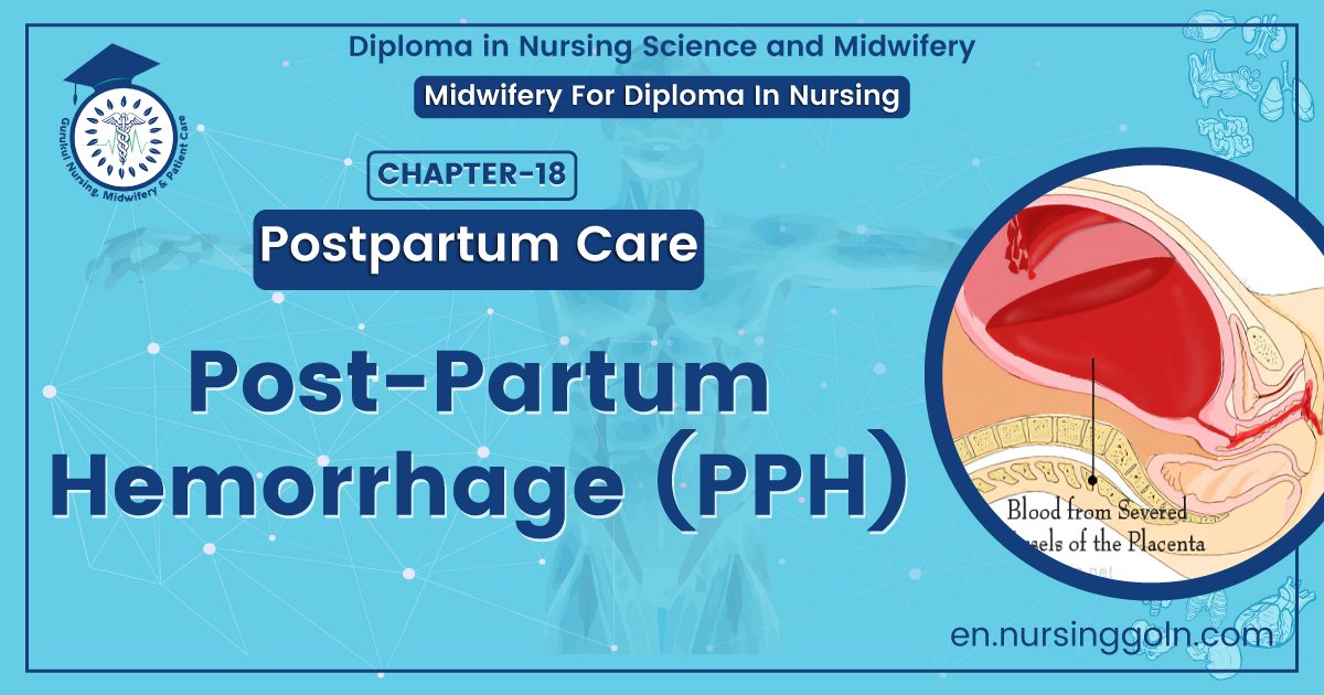
Post-Partum Hemorrhage (PPH)
Definition of PPH
Any amount of bleeding from or into genital tract following birth of the baby up to the end of the puerperium which adversely affects the general condition of the patient evidence by rise in pulse rate and falling blood pressure is called PPH.
Or
Postpartum bleeding or postpartum hemorrhage (PPH) is often defined as the loss of more than 500 ml or 1,000 ml of blood within the first 24 hours following childbirth.
Types of PPH
A. Primary PPH:
a) Excessive bleeding from or into the genital tract after birth within 24 hours is primary PPH. In the majority, hemorrhage occurs within 2 hours following delivery
b) Primary post-partum hemorrhage are mainly two types:
- 3rd stage Hemorrhage: Excessive bleeding before expulsion of placenta
- True PPH: Bleeding after expulsion of placenta (majority cases).
B. Secondary PPH: Excessive bleeding from or into the genital tract after 24 hours of delivery up to 6 weeks (the end of puerperium) is secondary PPH.
Causes of PPH (Post-Partum Hemorrhage)
| A. Causes of primary PPH: | 1. Uterine atony (80%): Predisposing causes are olde
2. Trauma (20%) |
| B. Causes of secondary hemorrhage (Hge): | 1. Retained bits of placenta or membrane – commonest 2. Infection 3. Sub involution 4. Secondary hemorrhage from caesarian section wound (uterus) 5. Choriocarcinoma 6. Cervical carcinoma omment 7. Regeneration of uterine fibroid 8. Polyplan |
A. Clinical features of PPH
a) Symptoms: Bleeding per vagina within 24 hours of delivery of fetus. It is usually visible but rarely may it be concealed.vooe som
b) Signs:
1. Signs of hemorrhage and shock.
2. Per abdominal examination-
- Uterus is soft and flaccid. Height at higher level.
- In traumatic hemorrhage-Uterus contracted,
3.. Per vaginal examination:
- Bleeding either copious or slow oozing or intermittent gushes occurs.
- Placenta delivered, not delivered or partially delivered. Injury at the birth canal may present.
B. Management of primary PPH
1. Shout for help to mobilize all the staffs.
2. Measures to stop the bleeding and to resuscitate simultaneously
3. A quick evaluation of the patient
- Whether patient is haemodynamically stable or not.
- Whether uterus is contracted or not.
- Whether a cord is found hanging or not.
4. To stop bleeding – Inj. Ergometrin 2 ampules IM or IV-if IV fluid remains (for sustained contraction).
5. Resuscitation of the patient:
- IV cannula should be inseted.
- Blood is sent for grouping and cross matching.
- IV fluid infusion rich in oxytocin. (4 ampules in 1000 ml. Hartmans solution is best, if it is not found DNS).
- Blood transfusion.
- antibiotics
- Catheterization
6. Gentle uterine massage by left hand if uterus is flabby.
7. Placenta is removed by controlled cord traction.
8. If placenta is not separated then manual removal of placenta under G/A
9. If bleeding is not stopped
- Bimanual compression of uterus or
- Aortic compression
10. If bleeding continues – Coagulopathy must be excluded to quitollinen sitesind
11. If bleeding continues – Patient is taken to the OT followed by
- Condom catheter: If normal vaginal delivary.
- B-lynch stitch, if C/S.
12. If bleeding continues:
- If family incomplete: Hysterectomy.
- If incomplete:
- Uterine arterial ligation.
- Ligation of anterior division internal life artery (rare).
13. If morbid adhesion of placenta is detected:
- Hysterectomy is the Rx of choice.
- If fertility is very important.
- Keep the placenta with cord in situ.
- Broad spectrum antibiotics.
- Autolysis of placenta with sloughing of the placenta.
14. After gentle massage of the uterus, if the uterus contracted, PPH is due to laceration of genital tract and then laceration should be repaired.
Prevention of PPH
A. Antenatal:
- Improvement of the health status of the mother and to keep the Hb level normal (>10 mg/dl).
- High risk patient are to be screened and delivered in a well-equipped hospital.
- Blood grouping should be done for all women.
B. Intra-natal:
- Slow delivery of the baby.
- Expert obstetric anesthetist in needed when delivery is conducted under G/A.
- During C/S spontaneous separation of placenta reduces blood loss.
- Active management of 3rd stage should be a routine.
- Examination of placenta and membrane should be a routine.
- In all cases of induced or accelerated labour by oxytocin, the infusion should be continued for at least one hour after delivery.
- Exploration of utero vaginal canal for evidence of trauma following difficult labour or instrumental delivery.
- To observe the patient for about two hours after delivery and if the uterus remains hard and contracted only then she should be send to the ward.
Complication of PPH
1. Bleeding may be very rapid.
2. Puerperal anemia and morbidity.
3. Puerperal sepsis.
4. Pulmonary embolism.
5. Fear for further pregnancies.
6. Hemorrhage is terrifying for the mother.
Management of secondary PPH

A. Clinical features:
- P/V bleeding-bright red and of varying amount.
- Anemia
- Evidence of sepsis
- P/V exam: May reveals sub involution of uterus with patulous cervical OS.
B. Investigation:
- Blood: TC, DC, ESR, Hb%
- Blood grouping, Rh typing and c Cross matching.
- Blood culture
- USG of pelvic organs.
C. Treatment:
1. General Rx:
- Bed rest
- Fluid
- Blood transfusion, if necessary.
- Cauterization
- Ergometrine 0.5 mg IM
- Broad spectrum antibiotic + Metronidazole.
2. Active Rx:
Exploration of the uterine cavity under G/A if retained products are seen. The suction curette may also be used.
Bleeding sometimes persists after evacuation. Occasionally packing uterus and even hysterectomy are required.