Physiology of Hearing-The course is designed for the basic understanding of anatomical structures and physiological functions of human body, musculoskeletal system, digestive system, respiratory system; cardiovascular system; urinary system, endocrine system, reproductive system, nervous system, hematologic system, sensory organs, integumentary system, and immune system.The aim of the course is to acquire knowledge and skills regarding anatomy and physiology.
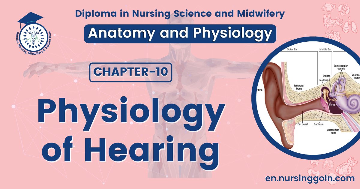
Physiology of Hearing
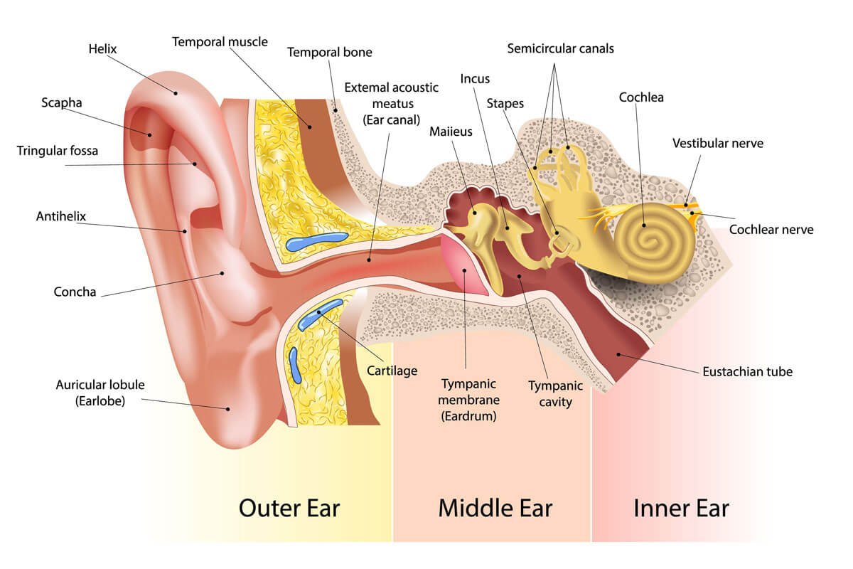
The events involved in stimulation of hair cells by sound waves are as follows:
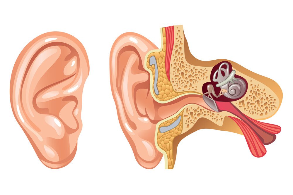
1. The auricle directs sound waves into the external auditory canal.
2. Sound waves striking the eardrum cause it to vibrate. The distance and speed of its movement depend on the intensity and frequency of the sound waves. More intense (louder) sounds produce larger vibrations. The eardrum vibrates slowly in response to low-frequency (low-pitched) sounds and rapidly in response to high-frequency (highpitched) sounds.
3. The central area of the eardrum connects to the malleus, which also starts to vibrate. The vibration is transmitted from the malleus to the incus and then to the stapes.
4. As the stapes moves back and forth, it pushes the oval window in and out.
5. The movement of the oval window sets up fluid pressure waves in the perilymph of the cochlea. As the oval window bulges inward, it pushes on the perilymph of the scala vestibuli.
6. The fluid pressure waves are transmitted from the scala vestibuli to the scala tympani and eventually to the membrane covering the round window, causing it to bulge outward into the middle ear.
7. As the pressure waves deform the walls of the scala vestibuli and scala tympani, they also push the vestibular membrane back and forth, creating pressure waves in the endolymph inside the cochlear duct.
8. The pressure waves in the endolymph cause the basilar membrane to vibrate, which moves the hair cells of the spiral organ against the tectorial membrane. Bending of their hairs stimulates the hair cells to release neurotransmitter molecules at synapses with sensory neurons that are part of the vestibulocochlear (VIII) nerve. Then, the sensory neurons generate nerve impulses that conduct along the vestibulocochlear (VIII) nerve.
(Ref:- J. Tortora, 8th edition, P-317)
Mechanism of hearing
Sound wave → strike the tympanic membrane → the wave transformed by tympanic membrane & auditory ossicles into movements of the foot plate of the stapes → this movements set up waves in the fluid of inner ear → the action of the waves on the organ of Corti, generates action potentials in the nerve fiber → signals go to the dorsal & ventral cochlear nuclei of medulla → then to superior olivary nucleus → and then to lateral lemniscus → pass through medial geniculate nucleus → finally to auditory cortex via auditory radiation (sound is audible).