Review of Anatomy & Physiology of Musculoskeletal System- An orthopedic nurse is a nurse who specializes in treating patients with bone, limb, or musculoskeletal disorders. Nonetheless, because orthopedics and trauma typically follow one another, head injuries and infected wounds are frequently treated by orthopedic nurses.
Ensuring that patients receive the proper pre-and post-operative care following surgery is the responsibility of an orthopedic nurse. They play a critical role in the effort to return patients to baseline before admission. Early detection of complications following surgery, including sepsis, compartment syndrome, and site infections, falls under the purview of orthopedic nurses.
Review of Anatomy & Physiology of Musculoskeletal System
The two primary subfields of orthopedic nursing are unplanned and elective surgery. Thus, elective surgery is when a patient’s quality of life is improved by a limb replacement, such as a hip or knee replacement, that has been detected during routine checkups.Some patients suffer an unanticipated injury, like a fractured hip, maybe while out for a walk. Within 48 hours of arriving at A&E, they will probably be clerked, admitted to an orthopedic ward, and undergo surgery.
Cell
Human cell:
Cell is the structural and functional unit of human body.
Cells from tissue,
Tissues from organ,
Organs from system,
Systems from Human body.
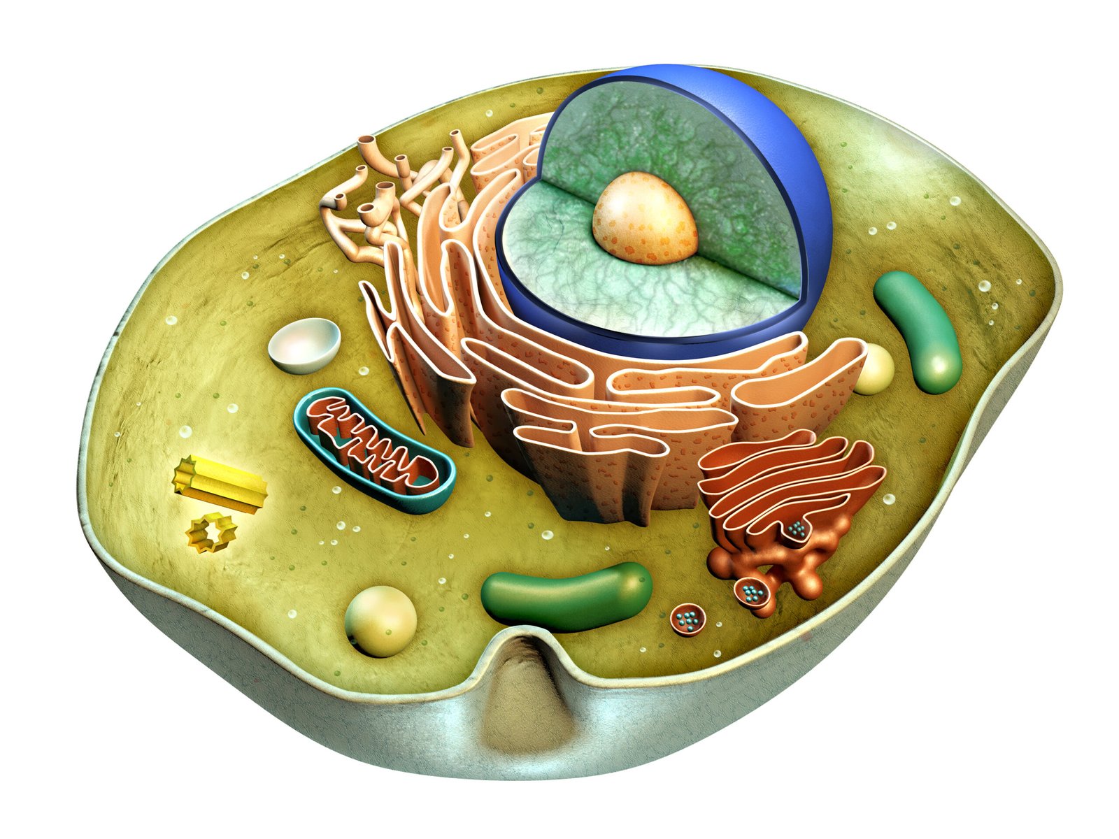
Parts of Cells :
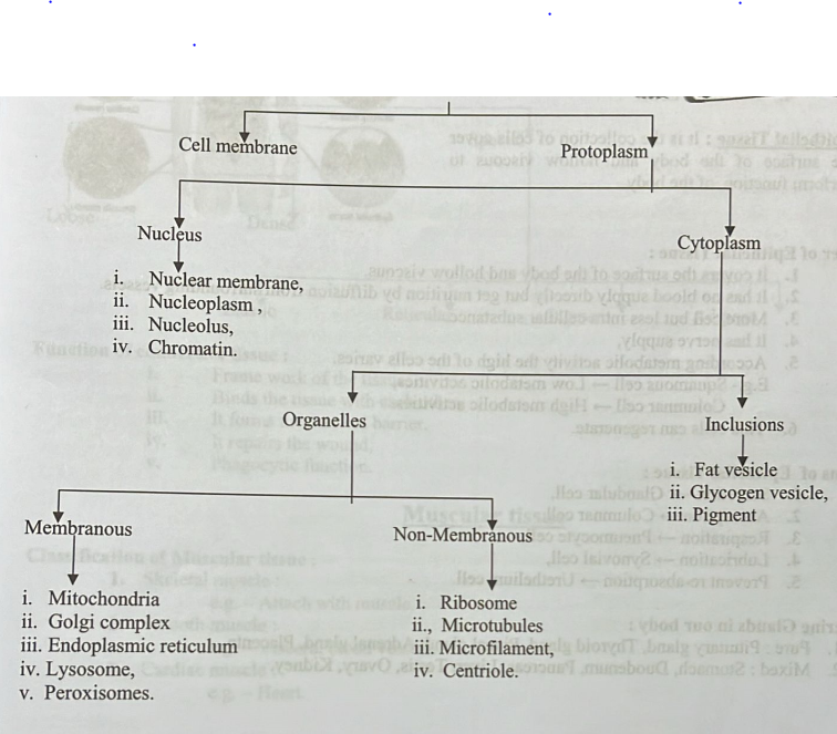
Function of cell :
Tissue
Definition of Tissue:
Collection of similar cells to perform functions of the body.
Classification of Tissue :
1. Epithelial Tissue: It is the collection of cells cover the surface of the body and hollow viscous to perform function of the body.
Character of Epithelial Tissue :
1. It covers the surface of the body and hollow viscous.
2. It has no blood supply directly but get nutrition by diffusion from surrounding vessels.
3. More cell but less intra cellular substance.
4. It has nerve supply,
5. According metabolic activity the high of the cells varies.
E.g.- Squamous cell Low metabolic activities,
Columnar cell High metabolic activities.
6. This cells can regenerate.
Functions of Epithelial Tissue:
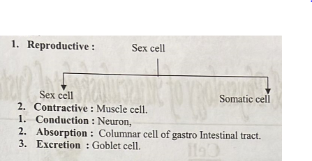
1. Secretion Glandular cell,
2. Absorption → Columnar cell,
3. Respiration → Pneumocyte cell,
4. Lubrication → Synovial cell,
5. Prevent re-absorption → Urethelium cell.
Endocrine Glands in our body:
1. Pure: Pituitary gland, Thyroid gland, Pineal gland, Adrenal gland, Placenta
2. Mixed: Stomach, Duodenum, Pancreas, Liver, Testis, Ovary, Kidney.
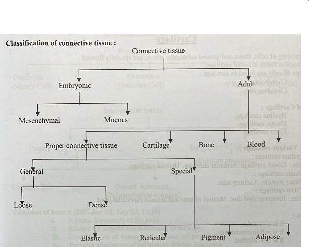
Connective tissue
Definition: It may consists of cell fibers and ground substances to binds the tissue with each other.
Characters:
i. Intercellular substance more but less number of cell.
ii. It binds the cells.
Function of connective tissue:
i. Frame work of the tissue,
ii. Binds the tissue with each other,
ⅲ. It forms mechanical barrier,
iv. It repairs the wound,
V. Phagocytic function.
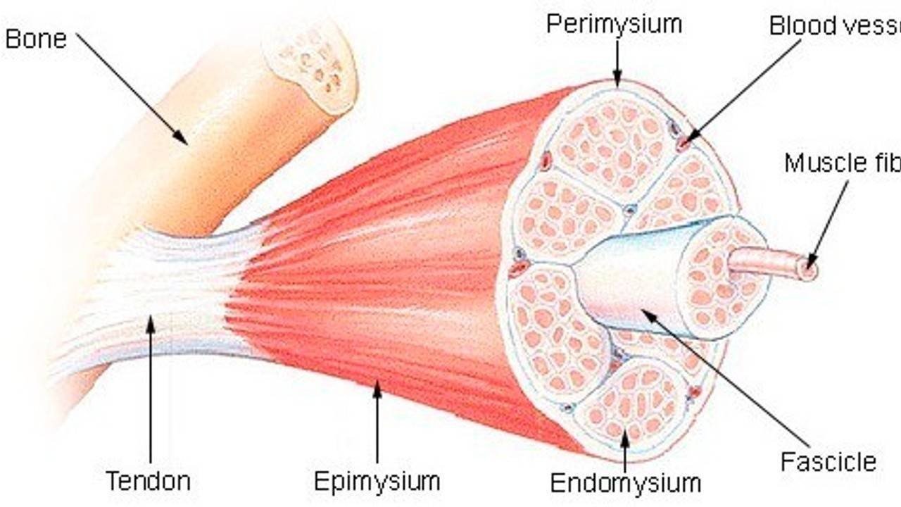
Muscular tissue
Classification of Muscular tissue:
1. Skeletal muscle: e.g. Attach with muscle.
2. Smooth muscle: e.g. – Viscera, blood vessels.
3. Cardiac muscle: e.g. – Heart.
Fibers
1. Collagen fiber:
Location: Tendon, ligaments.
2. Elastic fiber:
Location: Ligamentum flavum.
3. Reticular fiber:
Location: Lymphoid tissue, spleen bone marrow.
Cartilage
Definition: Consisting of cells, fibers and ground substances whose are specially formed connective tissue is called cartilage.
There are two type of cells are found in cartilage:
i. Chondroblast,
ii. Chondrocyte.
Classification of Cartilage:
i. Hyaline cartilage,
ii. Elastic cartilage,
iii.Fibro cartilage.
Various site of Various cartilage:
i. Hyaline cartilage:
Site: Costal cartilage, Articular cartilage, Thyroid cartilage.
ii. Elastic cartilage:
Site: Auricle, Auditory tube.
iii. Fibro cartilage:
Site: Inbtervertibral disc, Menisci of knee and acronio clavicullar joint.
Characteristics:
i. It is a vascular,
ii. It is soft in consistency,
iii.It is not calcified.
Functions:
i.It forms the skeleton of the body,
ii. They allow certain amount of movement,
iii.They acts as shock absorber,
iv. They protect the friction.
Different between Bone and Cartilage:
| Points | Bone | Cartilage |
| 1. Consistence | Hard | Soft |
| 2. Blood supply | Vascular | Avascular |
| 3. Nerve supply | Present | Absent |
| 4. Haversian system | Present | Absent |
| 5. Covering | Periosteum | Perichondrium |
Bone
Definition: Bone is specialized hard connective tissue for its mineralization.
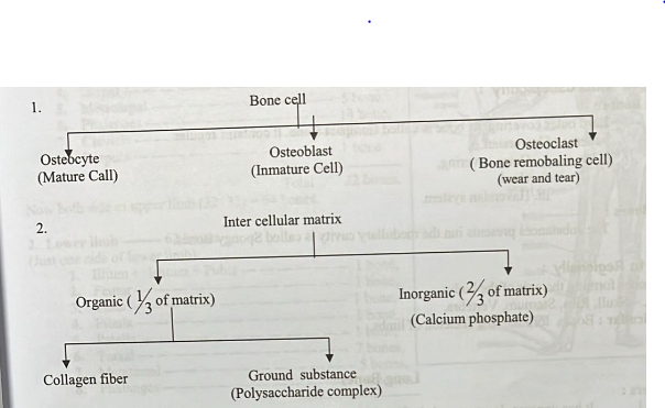
Composition of Bone:
Bone composed by:
1. Bone cell.
2. Inter cellular matrix.
Function of bone:
i. It forms framework of the body.
ii. It helps for support and movement of the body.
iii. Factory house of hemopoiesis (Blood cell produce).
iv. Provides attachment of the muscle, tendon, ligaments.
V.Protection of the viscera / organ.
vi. Store house of calcium (about 97% of body calcium)
Classification of Bone:
According to Morphologically:
1. Long Bones:
a. Typical:
i. Upper limb: Humerus, Radious, Ulna.
ii. Lower limb: Femur, Tibia, Fibula,
b. Miniature:
e.g. – Metacarpal. Metatarsal, Phalanges.
c. Modified:
e.g.- Clabicle, Body of Vertebra.
2 . Short Bones:
e.g. Carpals, Tarsals.
3. Flat Bones:
e.g. – Skull bones, Scapulla, Sternum, Ribs.
4. Irregular Bones:
e.g.- Hip bone, Vertebra, Base of the skull.
5. Pneumatice Bones :
e.g. – Maxillae, Sphenoid, Ethmoid.
6. Sasamoid Bones:
e.g. – Patella, Pisiform.
According to Macroscopically :
1. Compact Bones:
Hard outer covering of bone is called Compact Bone. It contains regular patterns of haversian.
It has:
i. Outer covering,
ii. Hard,
iii. Haversian system.
2. Spongy Bones:
The substances presents iun the medullary cavity is called Spongy Bones.
According to Regionally :
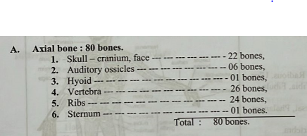
1. Axial: Bones forming the exis of the body. e.g. – Skull, Ribs, Sternum, Vertebra.
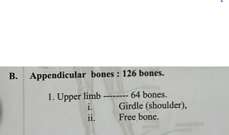
2. Appendicullar: Bones forming the skeleton of limbs
Long Bones
Characters:
Long Bones
i. It has upperen, loweren and shaft,
ii. It lies verticallay,
iii. It has outer compact and inner medullary cavity,
iv. Its ossification intracartelagenous,
V. It transmit body weight.

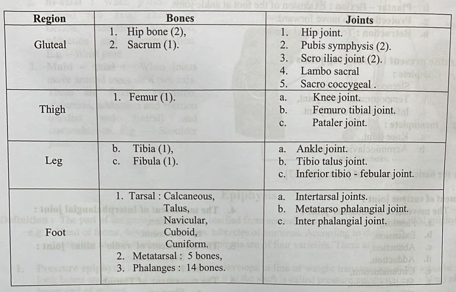
Number of bone :
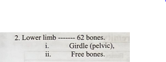
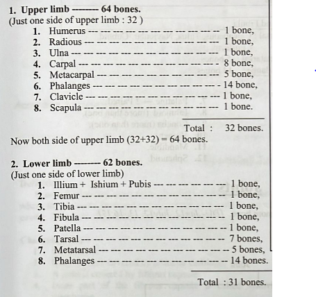
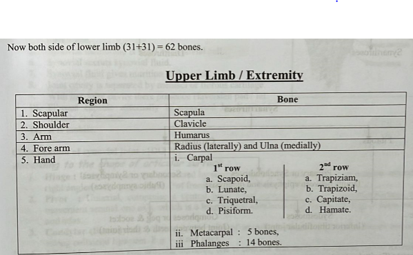
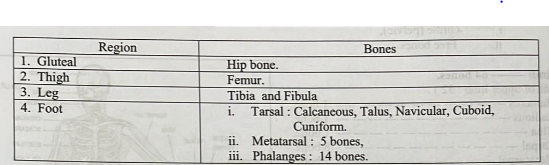
Lower Limb / Extremity
Bones of the skull :
1. Frontal bone,
2. Parietal bone 2 Paired,
3. Occipital bone,
4. Temporal bone → 2 Paired,
5. Zygomatic bone 2 Paired,
6. Maxilla→ 2 Paired,
7. Palatine 2 Paired,
8. Ethmoid (more than one),
10. Vomer,
9. Concha (more than one),
11. Mandible,
12. Sphenoid.
Joints 
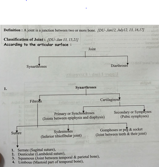
5. Plane (Joint between palatine process of maxilla),
6. Wedge & groove or schindylesis (Joint between rostram of sphenoid & vomer).
Diarthroses
1. Hinge (Humero- ulnar joint),
2. Pivot (Proximal radio – ulnar joint),
3. Condylar (Knee joint),
4. Ellipsoid (Sternoclavicular joint),
5. Saddle (Radio – carpal joint),
6. Ball & socket (Hip joint),
7. Plane (Sacroiliac joint)
According to movement :
1. Freely movement (Synovial joint),
2. Less mobile (Secondary cartilaginous joint),
3. No mobile/Immobile (Fibrous, primary cartilaginous joint.
Synovial Joint
Definition:
Synovial joints are connections between skeletal components where the elements involved are separated by a narrow articular cavity. All joints of the limbs are synovial joints.
Characteristics:
1. Connection.
2. Bony surface covered by articular cartilage.
3. A joint is covered by fibrous capsule.
4. Inner part of the fibrous capsule is called synovial membrane.
5. Synovial membrane is lined by synovial cell.
6. Synovial secrats synovial fluid. 7. Synovial fluid gives nutrition to the joint.
8. Joint cavity is separated by menisci or fibrous cartilage.
9. With in a joint cavity there presence Haversian pad/fat.
10. Fibrous capsule externally is supported by ligament, muscle and tendon.
11. It is mobile.
Classification of synovial joint:
According to the shape of articular surface:
1. Hinge: Uniaxial bones are connected by strong collateral ligaments. [Allow movement around one axis atun right angles to the bones (have l’of freedom)]. E.g. Humero- ulner joint.
2. Pivot: Uniaxial, compreise a central bony pivot surrounded by an osteo-ligamentous ring [Allow movement around one axis, which is a longitudinal axis(have 1º of freedom)]. E.g. Joint between dense mogrand atlas.
3. Condylar (Bicondylar): Uniaxial, two pairs of articular surfaces are presence. Bones are connected by strong collateral ligament. E.g. Knee joint.
4. Elipsoid: Bi-axial, two articular surfaces, one is concave and another is convex [allow movement in two
directions at right angles to each other (have l’of freedom)]. E.g. Radio carpal joint.
5. Saddle: Bi-axial saddle shaped bones fits into a socket [Allow movement around two horijontal axis at right angles to each other (have 2° of freedom)]. E.g. 1 carp metacarpal joint.
6. Ball and socket: Multiaxial here ball shaped head of bone fits into concave socket of another bone. [Allow movement in many directions (have 3° of freedom)]. E.g. – Shoulder joint.
7. Plane: Articular surfaces are flate, only gliding movement is possible. E.g. – Secroiliac joint.
According to the number of articular surface:
1. Simple: Possesses two articular surface. E.g. – Hip joint.
2. Compound: Possesses more than two articular surface. E.g.- Wrist joint.
3. Complex: Possesses more than two articular surface and the joint cavity is again divided into two by meniscus. E.g. – Knee joint.
According to the axis of movement:
1. Uniaxial : When joints move inlol Inte around one axis. This movement is flexion. E.g. – Elbow joint.
2. Bi-axial: When joints move around two axis. These are flexion, extension, abduction, adduction and surcumduction. E.g. – Wrist joint.
3 . Multi axial: When joints
move around more than two axis. These are flexion, extension, abduction, adduction and rotation (medial and lateral) and circumduction. E.g. Shoulder joint.
Epiphysis
Definition: The part of the growing long bone ossified from secondary ossification centre is called epiphysis. e.g. Head of femur, lower end of radius, tubercles of humerus. According to their activities as well as phylogeny the epiphysis are of four varieties. There are:
1. Pressure epiphysis: When the epiphysis develops in line of weight transmission at the articular ends of long bones and transmit body weight from it to the shaft is called pressure epiphysis. e.g. – Head of femur, lower end of radious.
2. Truction epiphysis: It is non articular, develops at the sites of attachment of ce rtain muscles, which exertraction (pulling action) on it. It ossified from independent ossification centre and generally appears at puberty. e.g. Greater and lesser trochanter of fumur, tubercles of humerus.
3. Atavistic epiphysis: It is the fusion of two epiphysis ossified from individual ossification centre (Phylogenetically they are separate bones). In human body there are only two. e.g. – Coracoids process of scapula and posterior tubercle of the talus.
4. Aberrant epiphysis: Only the first metacarpal bone possess only one epiphysis at its proximal end, other have only one epiphysis at their distant end when an epiphysis developed at the proximal end, than it is known as aberrant epiphysis.
Joint movements:
The movement permitted in joints are as follows.
1. Gliding movements: Surface glides over another (some carpal & tarsal articulation)
2.Angular movement :
a. Flextion: To bend or to make an angle.
b. Extension: To stretch out or to straighten.
c. Abduction: To draw away laterally from the median plane of the body.
d. Adduction: To draw towards the medial plane of the body,
e. Circumfusion: To perform the movements of flexion, abduction, extension and adduction sequence.
3. Rotatory movement: To rotate is to turn or revolve on a long axis, as the arm at shoulder joint.
a. Medial rotation,
b. Lateral rotation.
4. Special movements :
a. Supination: To rotate the forearm laterally which terms the plam forwards.
b. Pronation: to rotate the fore arm medially which brings the dorsum of hand 2forwards.
c. Inversion: Medial border of the foot is elevated so the sole faces laterally.
d Dorsiflexion : Flexion of the foot at ankle joint.
e.Evertion: Lateral border of the foot is elevated, so that the sole faces laterally.
f. Plantar-flexion : Extension of the foot at ankle joint.
g. Protection: To move forward.
h. Retraction: To move backwards.
Articular disc present in following joints:
1. Complete:
a. Sternoclavicular joint,
b. Temporomandicular joint,
c. Inferior radio-ulnar joint.
2. Incomplete:
a. Knee joint,
b. Acromioclavicular joint.
Movement of various joint:
1. The movement of shoulder joint :
a. Flextion,
b. Extension,
c. Abduction,
d. Adduction,
e. Circumduction,
f. Medial rotation,
g. Lateral rotation.
2. The movement of elbow joint :
a. Flextion,
b. Extension.
3. The movement of wrist joint:
a. Flextion,
b. Extension,
c. Abduction,
d. Adduction.
4. The movement of Interphalangial joint:
a. Flextion,
b. Extension,
5. The movements of radio- ulnar joint:
a. Supination,
b. Pronation.
6. The movement of Thumb:
a. Flextion,
b. Extension,
Abduction,
d. Adduction,
e. Circumduction,
The joint of upper limb :
The other name of bone is limb.
1. Sternoclavicular joint,
2. Acromioclavicolar joint,
3. Gleno-humeral joint (shoulder joind),
4. Elbo joint:
a. Humero ulnar loint,
b. Humero radial joint.
5. Superior radio-ulnar joint,
6. Inferior radio-ulnar joint,
7.Interosseus joint,
8. Wrist joint:
a. Radio carpal joint,
b. Inter carpal joint.
9. Carpometacarpal joint,
10. Metacarpo phalangial joint,
11. Inter phalangial joint.
Two fracture (#) is occoured in wrist joint-
1. Colles fracture (#),
2. Smith fracture (#).
Two fracture (#) is occoured in elbow joint –
1. Supra condylar fracture (#),
2.Dislocation fracture (#).
Lower/Inferior extrimity :