Salivary glands-The course is designed for the basic understanding of anatomical structures and physiological functions of human body, musculoskeletal system, digestive system, respiratory system; cardiovascular system; urinary system, endocrine system, reproductive system, nervous system, hematologic system, sensory organs, integumentary system, and immune system.The aim of the course is to acquire knowledge and skills regarding anatomy and physiology.
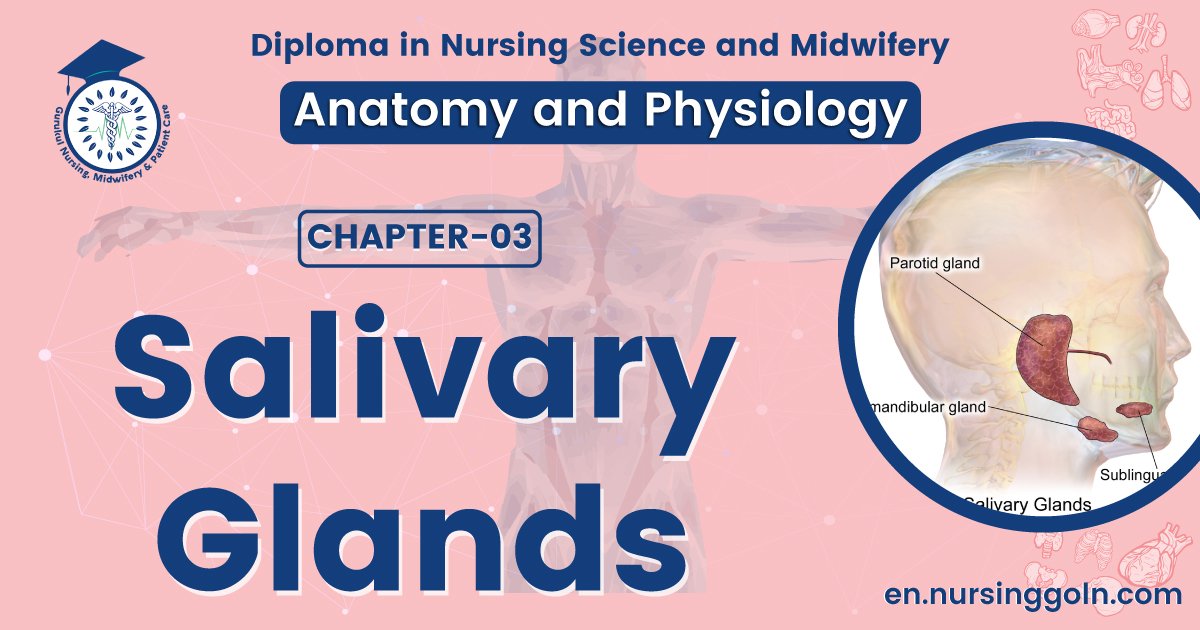
Salivary glands
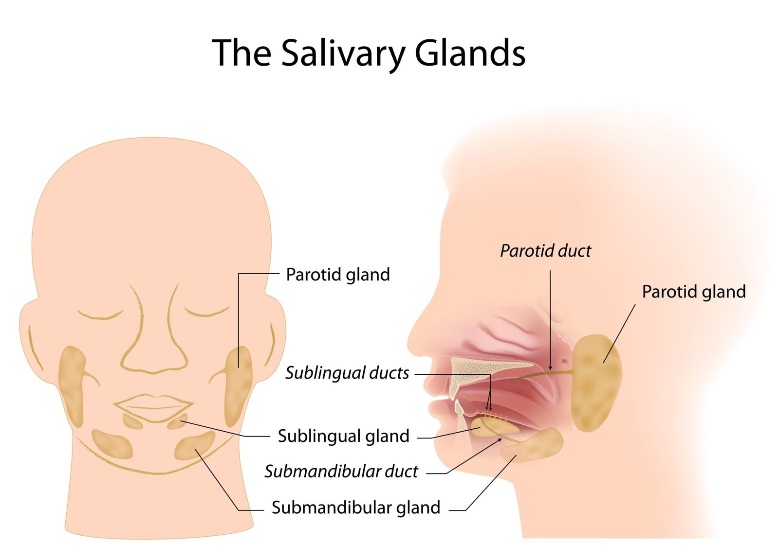
The salivary glands are accessory digestive glands that produce a secretion called saliva. Saliva functions as a solvent in cleansing teeth and dissolving food molecules so they can be tasted. Saliva. The three pairs of salivary glands are accessory organs of digestion that lie outside the mouth and release their secretions into ducts emptying into the oral cavity.
- The parotid glands are located inferior and anterior to the ears between the skin and the masseter muscle.
- The submandibular glands are found in the floor of the mouth; they are medial and partly inferior to the mandible.
- The sublingual glands are beneath the tongue and superior to the submandibular glands
Functions of saliva:
The fluid secreted by the salivary glands, called saliva, is composed of 99.5% water and 0.5% solutes.
- The water in saliva helps dissolve foods so they can be tasted and digestive reactions can begin.
- One of the solutes, the digestive enzyme salivary amylase, begins the digestion of starches in the mouth
- Mucus in saliva lubricates food so it can easily be swallowed.
- The enzyme lysozyme kills bacteria, thereby protecting the mouth’s mucous membrane from infection and the teeth from decay.
- Secretion of saliva, called salivation, which keeps the mucous membranes moist and lubricates the movements of the tongue and lips during speech. Sympathetic stimulation dominates during stress, resulting in dryness of the mouth.
(Ref-Guyton and Hall, Textbook of Medical Physiology, 12th ed)+ J. TORTORA, The essentials of anatomy and physiology, 8th edition, P-491)

Swallowing (deglutition), the movement of food from the mouth to the stomach, involves the mouth, pharynx, and esophagus and is helped by saliva and mucus. Swallowing is divided into three stages:
A. The voluntary stage,
B. Pharyngeal stage, and
C. Esophageal stage.
A. In the voluntary stage of swallowing, the bolus is forced to the back of the mouth cavity and into the oropharynx by the movement of the tongue upward and backward against the palate. With the passage of the bolus into the oropharynx, the involuntary pharyngeal stage of swallowing begins
B. In the pharyngeal stage Breathing is temporarily interrupted when the soft palate and uvula move upward to close off the nasopharynx, the epiglottis seals off the larynx, and the vocal cords come together. After the bolus passes through the oropharynx, the respiratory passageways reopen and breathing resumes. Once the upper esophageal sphincter relaxes, the bolus moves into the esophagus.
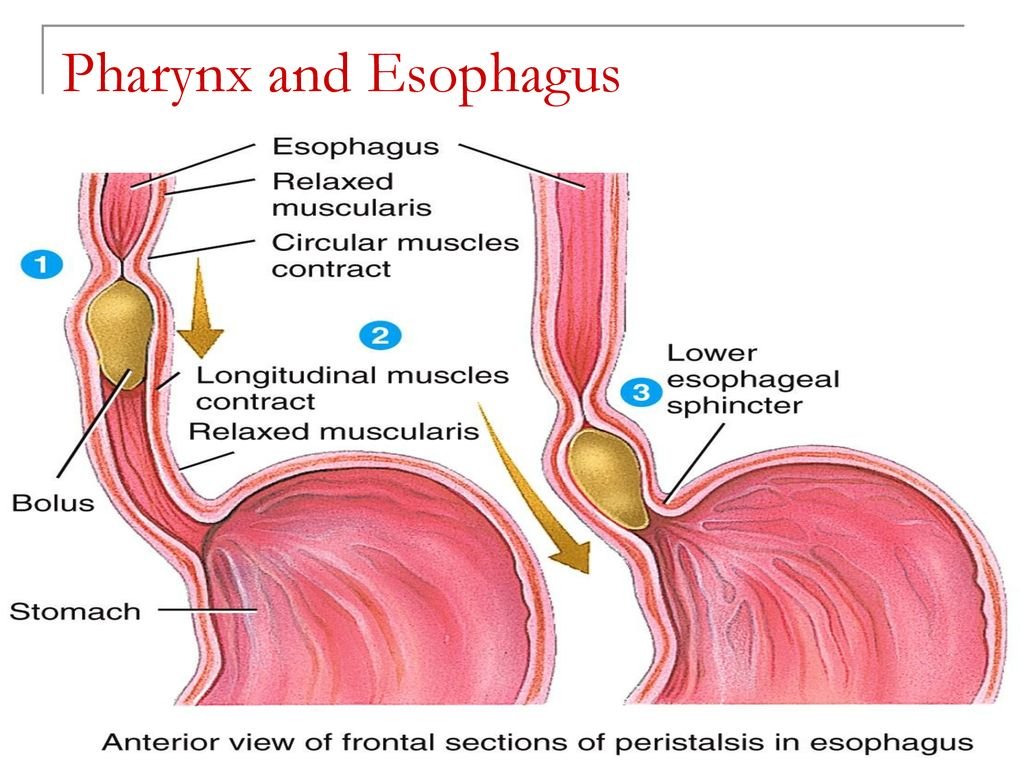
C. In the esophageal stage, food is pushed through the esophagus by a process called peristalsis:
1. The circular muscle fibers in the section of esophagus above the bolus contract. constricting the wall of the esophagus and squeezing the bolus downward.
2. Longitudinal muscle fibers around the bottom of the bolus contract, shortening the section of the esophagus below the bolus and pushing its walls outward.
3. After the bolus moves into the new section of the esophagus, the circular muscles above it contract, and the cycle repeats. The contractions move the bolus down the esophagus toward the stomach. As the bolus approaches the end of the esophagus, the lower esophageal sphincter relaxes and the bolus moves intothe stomach.
Read more: