
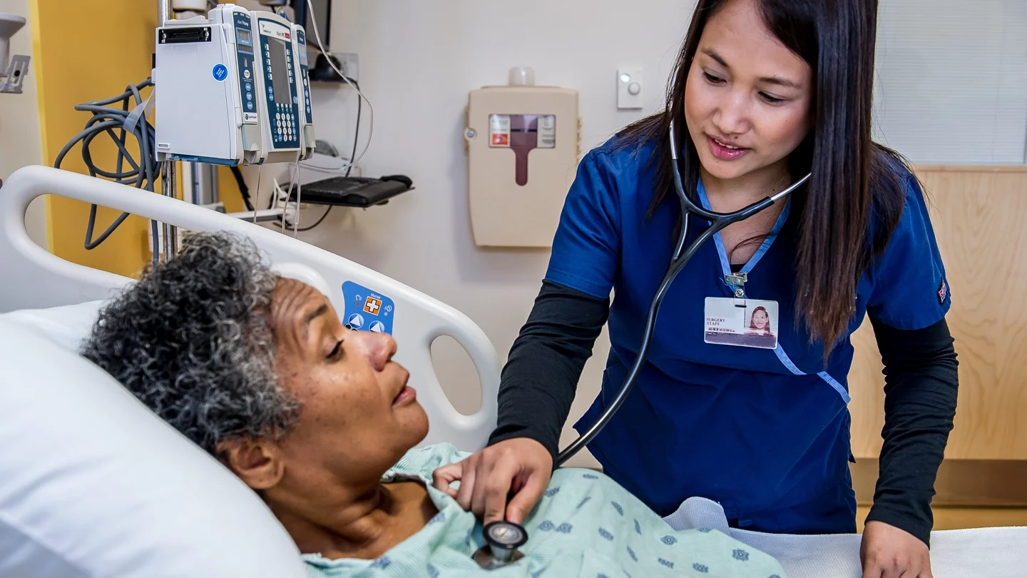
Today our topic of discussion is Sites for Hemodialysis.
Sites for Hemodialysis
Sites for Hemodialysis
- Subclavian vein catheterization: Using the Seldinger technique surgeon introduces the needle into subclavian vein then inserts a guidewire through the introducer needle and then removes the needle
- Using the guide wire thread 5″-12″ (12-30 cm) plastic or Teflon catheter (with a Y hub) into the patient’s vein
- Femoral vein catheterization: Using the Seldinger technique surgeon introduces the needle into the left or right femoral vein.
- Then insert a guide wire through introducer needle and remove the needle.
- Using the guide wire then thread a 5″-12″ plastic or Teflon catheter with a Y hub or two catheters one for inflow and another place about 1/2″ (1.3 cm) distal to the first for outflow
- Arteriovenous fistula: To create fistulas make an incision into the patient’s wrist or lower forearm then a small incision into the side of the artery and another side of a vein, then suture the edges of the incisions together to make a common opening 3-7 mm long (Fig. 29.116) Arteriovenous shunt: To create a shunt makes a incision in the patient’s wrist, lower forearm, or an ankle.
- Then insert 6″-10″ (15-25 cm) transparent silastic cannula into an artery and another into a vein. Finally, tunnel the cannulas out through a stab wound and join them with a piece of a Teflon tubing
- Arteriovenous graft: To create a graft makes an insertion in the patient’s forearm, upper arm or thigh.
- Then tunnel a natural or synthetic graft under the skin and suture the distal end to an artery and proximal end to a vein.
Mechanism of Hemodialysis (Fig. 29.117)
- In hemodialysis the blood flows from the patient to an external dialyzer (artificial kidney) through an arterial assess site.
- Inside the dialyzer the blood and the dialysate flow counter currently divided by a semipermeable membrane, the composition of the dialysate resembles normal extracellular fluid.
- Blood contains excess of specific solutes and dialysate contains electrolytes that may be at abnormal level in the patient’s bloodstream.
- The dialysate electrolyte composition can be raised or lowered depending on the need.
- Excretory function an electrolyte hemostasis area achieved by diffusion, the movement of molecule across the dialyzer’s semipermeable membrane from an area of higher solute concentration to an area of lower concentration.
- Water (solvent) crosses the membrane from the blood into a dialysate by ultrafiltration.
- This process removes excess water, waste products and other metabolites through osmotic pressure and hydrostatic pressure.

- Osmotic pressure is the movement of water across the semi permeable membrane from an area of lesser solute concentration to greater solute concentration.
- Hydrostatic pressure forces the water from the blood compartment into the dialysate compartment, cleaned from impurities and in excess water the purified blood returns to the body through a venous site.