Tape Worms – Basic microbiology, parasitology, and immunology; nature, reproduction, growth, and transmission of common microorganisms and parasites in Bangladesh; prevention including universal precaution and immunization, control, sterilization, and disinfection; and specimen collections and examination. Students will have an understanding of common organisms and parasites caused human diseases and acquire knowledge about the prevention and control of those organisms.
Tape Worms
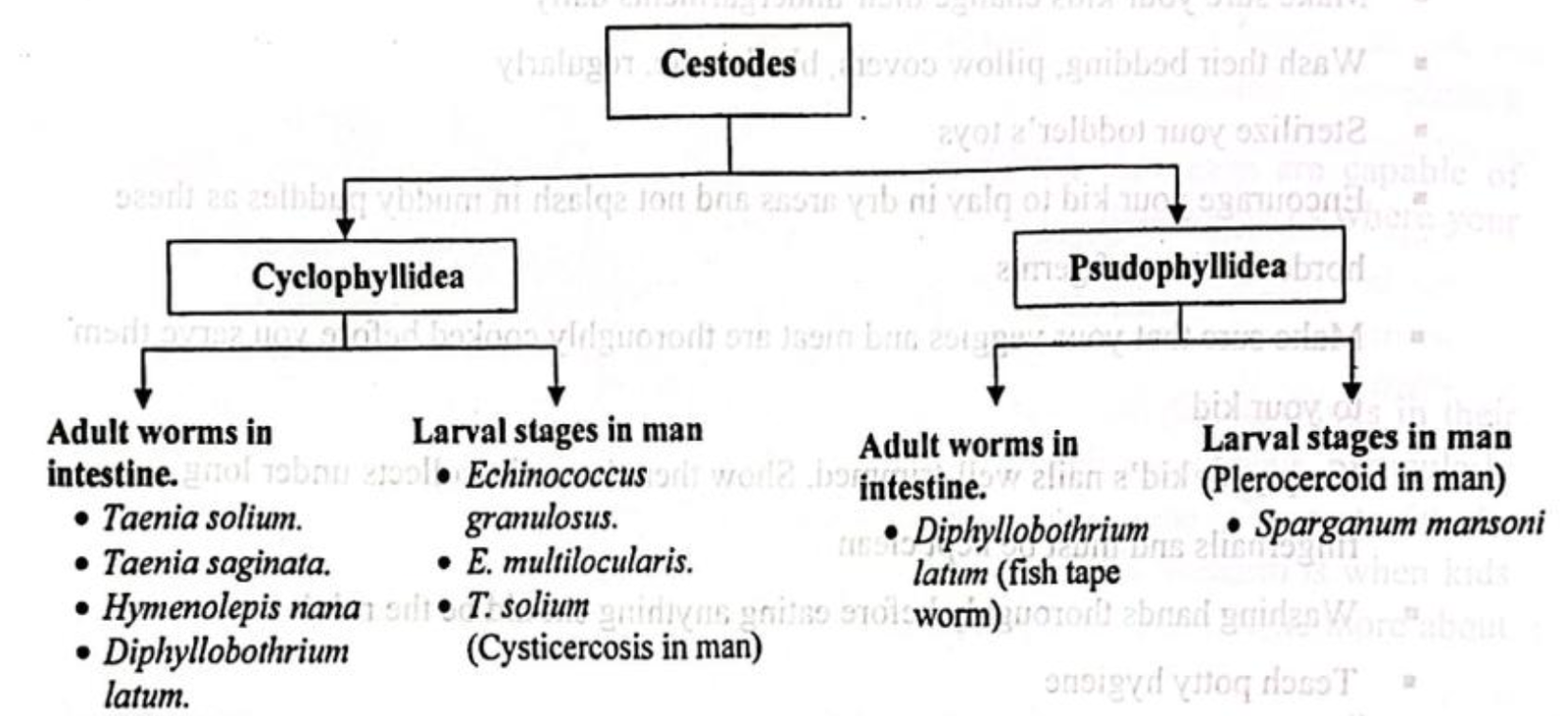
Classification of Tape Worms/ Cestode:
It can be classified in two ways
1. On the basis of habitat
2. On the basis of Systemic classification
Medically important cestodes are classified according to habitat. It is as follows –
According to habitat:
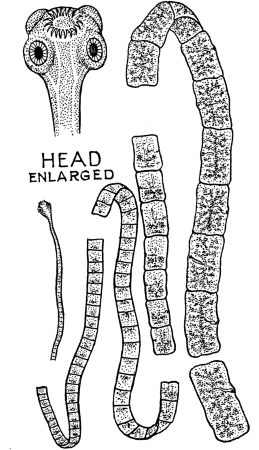
General Characters of Cestodes:
- Cestodes are segmented and tape like.
- Sizes vary from a few millimeters to several meters.
- Adult worms are found in the intestinal canal of man and animal.
- “Head” is provided with suckers (slit- like or cup-like) and sometimes with hooks which serve as organs of attachment.
- There are three regions in an adult worm: “head” (scolex), “neck” and a strobila (a body or trunk) consisting of a series of segments (proglottides).
- Sexes are not separate.
- Body cavity is absent.
- Alimentary canal is entirely absent.
- Excretory and nervous systems are present.
- Reproductive system is highly developed and complete in each segment.
(Thread Worm / Pin Worm)/ Enterobius Vermicularis
Life cycle of Enterobius vermicularis:
➤ Infective form: Embryonated egg containing tadpole like larva.
➤ Portal of entry: Alimentary canal (Faeco-oral route).
➤ Site of location: Caecum and vermiform appendix.
➤ Types of infection:
- Person to person
- Auto infection and
- Retrograde infection
➤ Life cycle:
- Host – Man is known host: No intermediate host is required
Stages
Newly laid egg in perianal skin, containing tadpole like larva → Completes its development within 24-36 hours in presence of O2 → Infection by ingestion of these eggs → In gut, egg shell dissolved by digestive juices → Larva escape in the small intestine → Develops into adolescent worm → sexually matures → Male fertilizes the female and dies → Gravid female migrates from small intestine to caecum and colon and vermiform appendix → Remain there until egg develops → Fertilized female then wander down the rectum and works it way out of the anus at night → Deposit egg in perianal skin→The cycle is repeated (Whole cycle requires 2 -4 weeks )
Mode of Transmission of Enterobius Vermicularis:
Ingestion of eggs by –
- Person to person
- Auto infection and
- Retrograde infection
Clinical Conditions Caused by Enterobius vermicularis:
- In heavy infection, abdominal pain, anal pruritus, pallor and dysentery.
- Perianal pruritus and an eczematous condition around the anus and perineum.
- Nocturnal enuresis (frequency of micturition).
- Vulvitis and vulvo-vaginitis in girls.
- Sometimes penetrate into the peritoneal cavity and cause salpingitis or even, peritonitis.
- UTI among young girls
- Sometimes inflammation of the vermiform appendix (in 2% of cases)
- Bilateral tubo-ovarian abscess
Laboratory Diagnosis of Enterobiasis:
➤ Principle: Microscopic identification of pinworm eggs in the perianal region is the evidence of infection. Eggs are rarely found in the feces, and the diagnosis is made by finding eggs on perianal swabs made of Scotch tape. The tape is pressed first onto the perianal region and then onto a microscope slide, and is examined microscopically. Perianal specimens are best obtained in the morning before bathing or defecation. Three specimens should be taken on consecutive days before pinworm infection is ruled out.
➤ Specimen collection: Perianal swab
➤ Microscopic examination:
- Coverslip preparation with saline.
- Demonstration of egg of E. vermicularis the perianal skin
➤ Macroscopic examination:
- Adult worm may be found in the perianal region at night or during passing stool.
➤ Non specific examination:
- Occult blood test-Often positive,
- Differential count of W.B.C.-may show eosinophilia