The acessory structures of the eye-The course is designed for the basic understanding of anatomical structures and physiological functions of human body, musculoskeletal system, digestive system, respiratory system; cardiovascular system; urinary system, endocrine system, reproductive system, nervous system, hematologic system, sensory organs, integumentary system, and immune system.The aim of the course is to acquire knowledge and skills regarding anatomy and physiology.
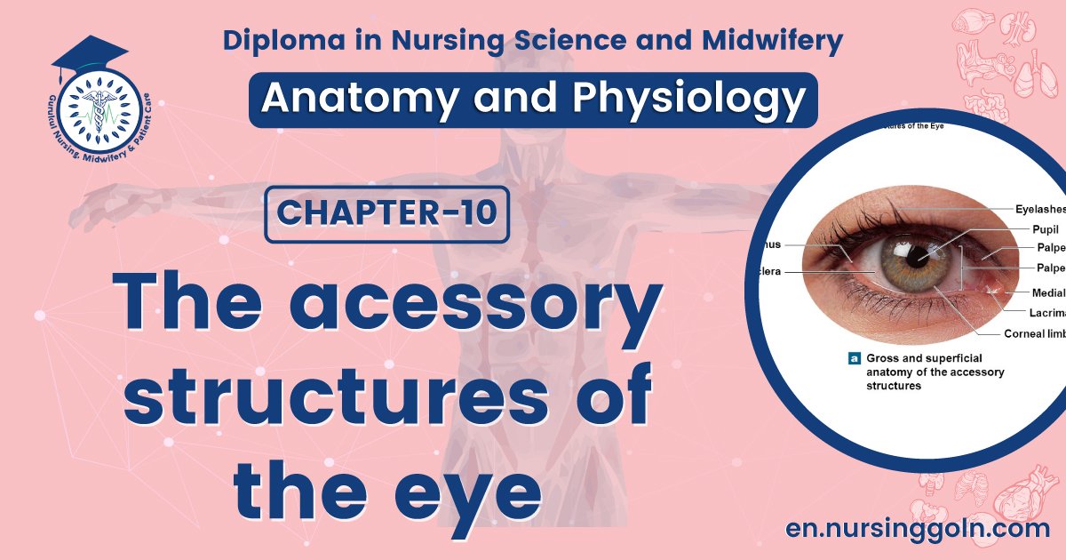
The acessory structures of the eye
More than half the sensory receptors in the human body are located in the eyes, and a large part of the cerebral cortex is devoted to processing visual information.
Accessory Structures of the Eye
The accessory structures of the eye are:-
- The eyebrows,
- Eyelashes,
- Eyelids,
- Extrinsic muscles that move the eyeballs, and
- Lacrimal (tear-producing) apparatus.
The eyebrows and eyelashes :-
The eyebrows and eyelashes help protect the eyeballs from foreign objects, perspiration, and direct rays of the sun.
The eyelids :-
The upper and lower eyelids shade the eyes during sleep, protect the eyes from excessive light and foreign objects, and spread lubricating secretions over the eyeballs (by blinking).
Extrinsic muscles of eye :-
Six extrinsic eye muscles cooperate to move each eyeball right, left, up, down, and diagonally: the superior rectus, inferior rectus, lateral rectus, medial rectus, superior oblique, and inferior oblique. Neurons in the brain stem and cerebellum coordinate and synchronize the movements of the eyes.
The lacrimal apparatus:-
The lacrimal apparatus (lacrima = tear) is a group of glands, ducts, canals, and sacs that produce and drain lacrimal fluid or tears. The right and left lacrimal glands are each about the size and shape of an almond.
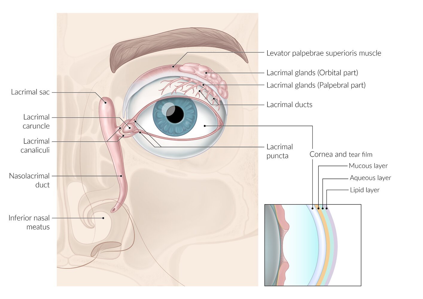
They secrete tears through the lacrimal ducts onto the surface of the upper eyelid. Tears then pass over the surface of the eyeball toward the nose into two lacrimal canals and a nasolacrimal duct, which allow the tears to drain into the nasal cavity.
Tears are a watery solution containing salts, some mucus, and a bacteria-killing enzyme called lysozyme. Tears clean, lubricate, and moisten the portion of the eyeball exposed to the air to prevent it from drying.