The humerus – The course is designed for the basic understanding of anatomical structures and physiological functions of human body, musculoskeletal system, digestive system, respiratory system; cardiovascular system; urinary system, endocrine system, reproductive system, nervous system, hematologic system, sensory organs, integumentary system, and immune system.
The aim of the course is to acquire knowledge and skills regarding anatomy and physiology.
The humerus
The Humerus
The humerus is the longest bone of the arm and is characterized by many distinct features that help to allow the upper extremity to move through a significant range of motion. The following landmarks are found on the humerus:
Head. A ball-shaped structure that articulates with the glenoid cavity.
Anatomical neck. Formed by a narrow constriction immediately distal to the head of the humerus.
Surgical neck. Lies distal to the anatomical neck and tubercles of the humerus. The axillary nerve and the posterior humeral circumflex artery course into the posterior compartment of the arm, deep to the surgical neck.
Greater and lesser tubercles. Enlarged areas for muscle attachments
Intertubercular (bicipital) groove. A deep sulcus between the greater and lesser tubercles, where the long head of the biceps brachii tendon courses en route to the supraglenoid tubercle
Radial (spiral) groove. A distinct groove on the posterior surface of the humerus, where the radial nerve and the deep brachial artery course.
Deltoid tuberosity. A large V-shaped protrusion on the lateral-surface of the humerus, midway along its length where the deltoid muscle attaches.
Lateral epicondyle. Located on the distal lateral end of the humerus and provides an attachment surface for the posterior forearm muscles (extensors).
Medial epicondyle. Located on the distal medial end of the-humerus and provides an attachment surface for the anterior forearm muscles (flexors).
Trochlea. Characterized by a pulley shape; it helps to guide the hinge joint. The trochlea of the- humerus articulates with the trochlear notch of the ulna.
Capitulum. Characterized by its oval, convex shape for articulation with the radial head.
Coronoid fossa. Located on the distal anterior surface of the-humerus, where the coronoid process of the ulna articulates.
Olecranon fossa. Located on the distal posterior surface of the-humerus, where the olecranon process of the ulna articulates.
(Ref: Guyton and Hall, Textbook of Medical Physiology, 12th ed)
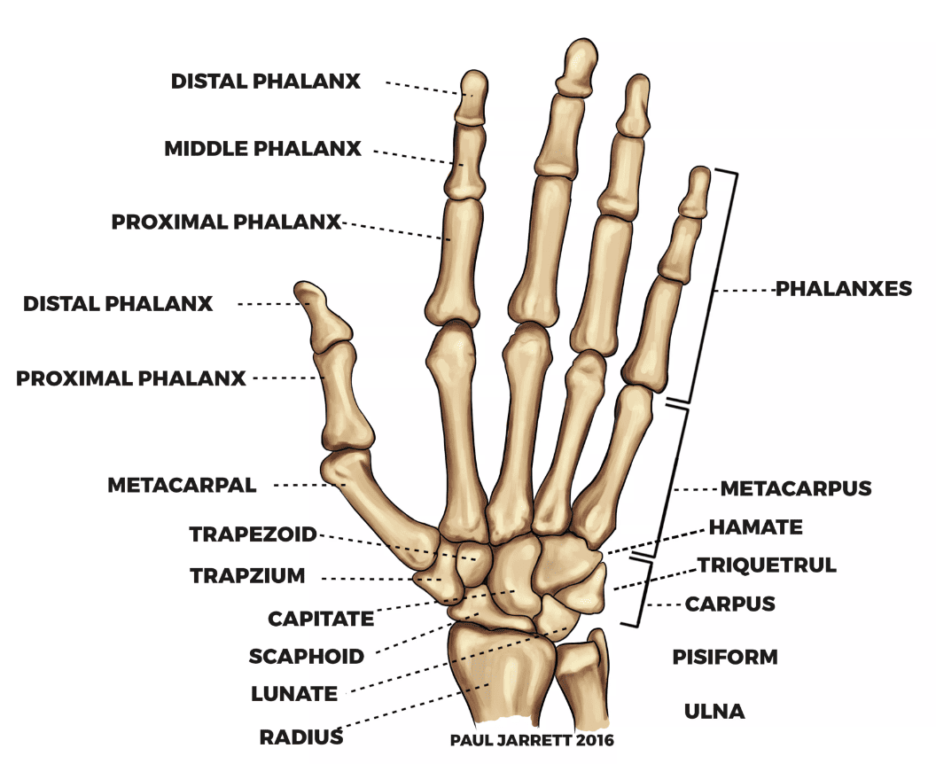
The skeleton of the hand consists of the carpals, metacarpals, and phalanges. And it consists of 27 bones in each hand. Both hands consist of total (27 X 2)= 54 bones.
One hand consists 27 bones are mentioned below as….
Carpals = 8 bones
- Scaphoid
- Lunate
- Triquetrum
- Pisiform
- Trapezium
- Trapezoid
- Capitate
- Hamate
Metacarpals = 5 bones
Phalanges = 14 bones
___________________________________
Total = 27 bones
Lower limbs consists of (30X2)=60 bones. So, each lower limbis composed of 30 bones: the femur in the thigh; the patella (kneecap); the tibia and fibula in the leg (the part of the lower limb between the knee and the ankle); and 7 tarsals (ankle bones), 5 metatarsals, and 14 phalanges (toes) in the foot.
Bones of the Lower limbs
Femur — –2
Patella—— 2
Fibula- —– 2
Tibia ——–2
Tarsals——14
Metatarsal–10
Phalanges –28
_____________________
Total = 60 bones