The lung-The course is designed for the basic understanding of anatomical structures and physiological functions of human body, musculoskeletal system, digestive system, respiratory system; cardiovascular system; urinary system, endocrine system, reproductive system, nervous system, hematologic system, sensory organs, integumentary system, and immune system.The aim of the course is to acquire knowledge and skills regarding anatomy and physiology.
The lung
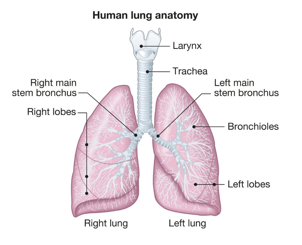
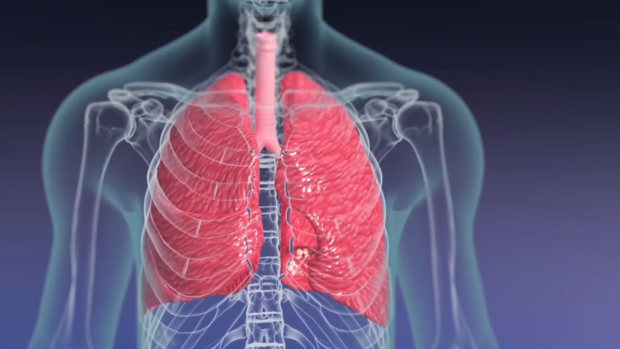
The lungs (= lightweights, because they float) are two spongy, cone-shaped organs in the thoracic cavity. There are two lungs which are separated from each other by the heart and other structures in the mediastinum.
Layers/covering of the lungs
The pleura/The pleural membrane is a double-layered serous membrane that encloses and protects each lung.
- The outer layer is attached to the wall of the thoracic cavity and diaphragm and is called the parietal pleura.
- The inner layer, the visceral pleura, is attached to the lungs.
- Between the visceral and parietal pleurae is a narrow space, the pleural cavity, which contains a lubricating fluid secreted by the membranes. This fluid reduces friction between the membranes, allowing them to slide easily over one another during breathing
External features
The lungs extend from the diaphragm to slightly above the clavicles and lie against the ribs.
- The broad bottom portion of each lung is its base,
- The narrow top portion is the apex
- The left lung has an indentation, the cardiac notch, in which the heart lies. Due to the space occupied by the heart, the left lung is about 10% smaller than the right lung
Surfaces:-
There are two surfaces..
- Costal surface
- Medial surface
Borders :-
There are three borders.
- Inferior border
- Anterior border
- Posterior border
Fissures and lobes :-
Deep grooves called fissures divide each lung into lobes.
The oblique fissure divides the left lung into two lobes
- Superior lobe and
- Inferior lobe.
The oblique and horizontal fissures divide the right lung into three lobes
- Superior lobe,
- Middle lobe, and
- Inferior lobe

Bronchus and it’s branches:-
- Each lobe receives its own secondary bronchus. Each lung lobe is divided into smaller segments that are supplied by a tertiary bronchus. The segments, in turn, are subdivided into many small compartments called lobules.
- Each lobule contains a lymphatic vessel, an arteriole, a venule, and a branch from a terminal bronchiole wrapped in elastic connective tissue.
- Terminal bronchioles subdivide into microscopic branches called respiratory bronchioles, which are lined by nonciliated simple cuboidal epithelium
- Respiratory bronchioles, in turn, subdivide into several alveolar ducts. The two or more alveoli that share a common opening to the alveolar duct are called alveolar sacs.
- Alveoli: An alveolus (plural is alveoli) is a cup-shaped outpouching of an alveolar sac. Many alveoli and alveolar sacs surround each alveolar duct. The walls of alveoli consist mainly of thin alveolar cells, which are simple squamous epithelial cells. They are the main sites of gas exchange.
Functions of the lungs
- The functions of the lungs is mainly the exchange of gases (O, and Co₂) between the atmosphere and blood in the body.
- The lungs possess several characteristics which protect against infection. The lung tract is lined by epithelia with hair-like projections called cilia that beat rhythmically and carry mucus. This mucociliary clearance is an important defence system against air-borne infection.
(Ref:- K. Indu, 1 ed P-223,224 + P.Evelyn, 16h,P-257-261 + J. Tortora, 8th ed, P-465 + Ross and wilson- 9 ed, P-249,251
Read more: