
The structure and functions of glands-The course is designed for the basic understanding of anatomical structures and physiological functions of human body, musculoskeletal system, digestive system, respiratory system; cardiovascular system; urinary system, endocrine system, reproductive system, nervous system, hematologic system, sensory organs, integumentary system, and immune system.The aim of the course is to acquire knowledge and skills regarding anatomy and physiology.

The structure and functions of glands
Accessory structures of the skin that develop from the epidermis of an embryo-hair, glands, and nails-perform vital functions. Hair and nails protect the body. Sweat glands help regulate body temperature.
Hair
Hairs, or pili, are present on most skin surfaces except the palms, palmar surfaces of the fingers, soles, and plantar surfaces of the toes. In adults, hair usually is most heavily distributed across the scalp, over the brows of the eyes, and around the external genitalia.
Genetic and hormonal influences largely determine the thickness and pattern of distribution of hair. Hair on the head guards the scalp from injury and the sun’s rays; eyebrows and eyelashes protect the eyes from foreign particles; and hair in the nostrils protects against inhaling insects and foreign particles.
Glands
Glands are single or groups of epithelial cells that secrete a substance. The glands associated with the skin include sebaceous, sudoriferous, and ceruminous glands.
Sebaceous Glands
Sebaceous glands (sebace greasy) or oil glands, with few exceptions, are connected to hair follicles. The secreting portions of the glands lie in the dermis and open into the hair follicles or directly onto a skin surface.
There are no sebaceous glands in the palms and soles. Sebaceous glands secrete an oily substance called sebum. Sebum keeps hair from drying out, prevents excessive evaporation of water from the skin, keeps the skin soft, and inhibits the growth of certain bacteria. Sebaceous gland activity increases during adolescence.
Nails
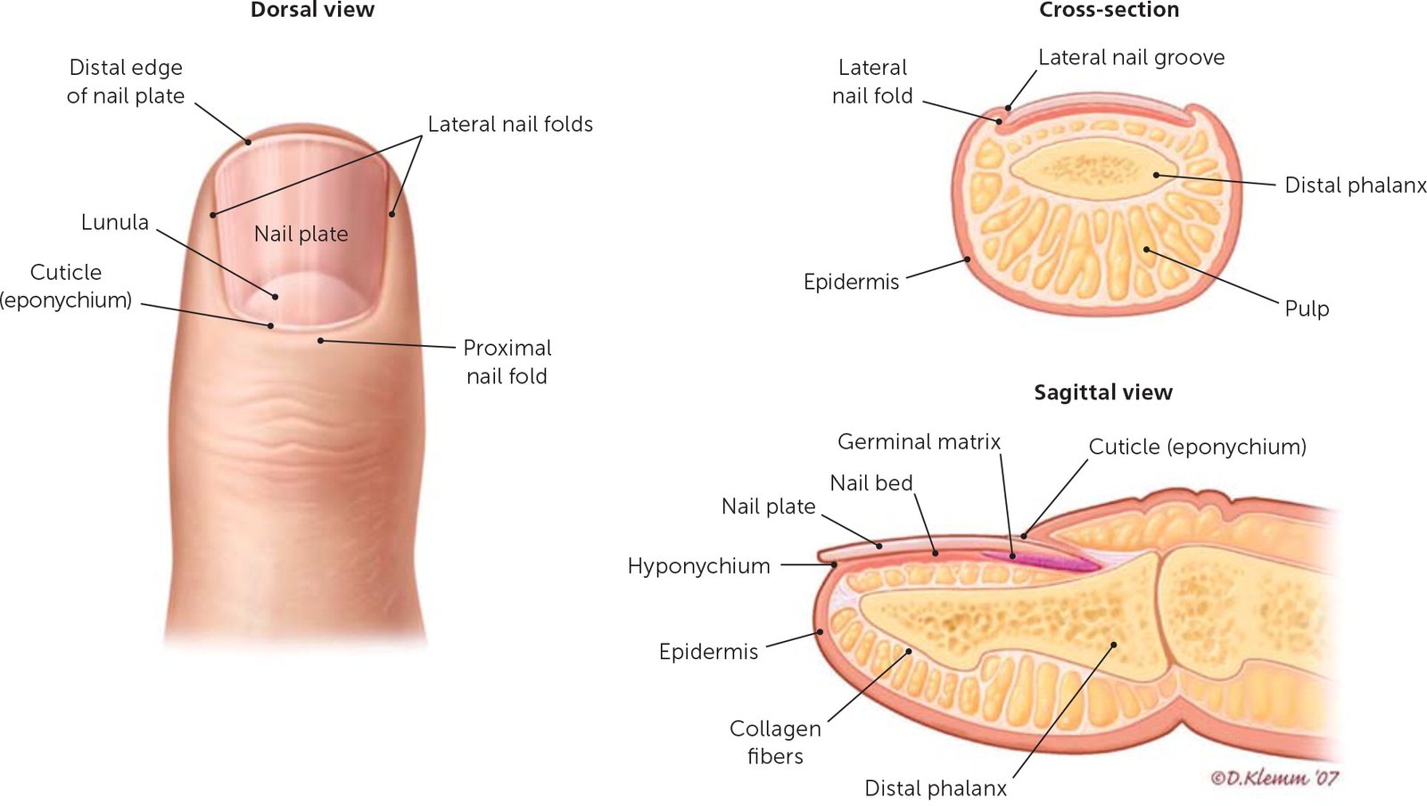
Nails are plates of tightly packed, hard, dead, keratinized cells of the epidermis. Each nail consists of a nail body, a free edge, and a nail root. The nail body is theportion of the nail that is visible; the free edge is the part of the body that extends past the end of the finger or toe; the nail root is the portion that is not visible.
Most of the nail body is pink because of the underlying blood capillaries. The whitish semilunar area near the nail root is called the lunula (little moon). It appears whitish because the vascular tissue underneath does not show through due to the thickened stratum basale in the area.
The proximal portion of the epithelium deep to the nail root is called the nail matrix. It is in this region that the superficial cells divide by mitosis to produce new nail cells. The average growth of fingernails is about 1 mm (0.04 inch) per week. The cuticle consists of stratum corneum.
Function of nail:
Nails help us grasp and manipulate small objects, provide protection to the ends of the fingers and toes, and allow us to scratch various parts of the body.