Trichomonas vaginalis – Basic microbiology, parasitology, and immunology; nature, reproduction, growth, and transmission of common microorganisms and parasites in Bangladesh; prevention including universal precaution and immunization, control, sterilization, and disinfection; and specimen collections and examination. Students will have an understanding of common organisms and parasites caused human diseases and acquire knowledge about the prevention and control of those organisms.
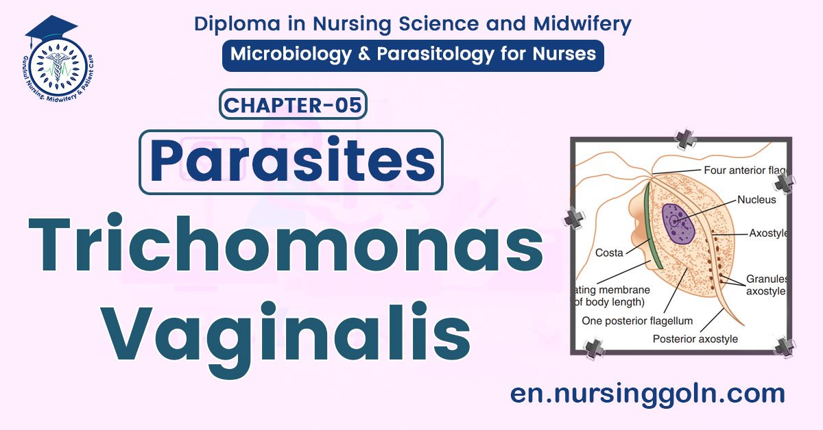
Trichomonas vaginalis
Trichomonas vaginalis causes Trichomoniasis.
Habitat:
- In the female genital tract (Primarily vagina).
- the urinary tract of female and male (Primarily prostate).
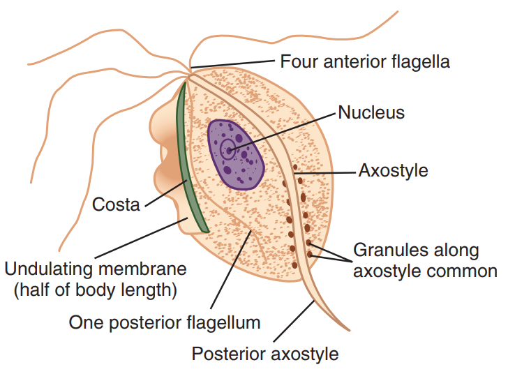
Morphology:
- Pear shaped.
- Only trophozoite form and no cystic stage.
- A central nucleus and four anterior flagella
- It has an undulating membrane that extends about two-thirds of its length.
Mode of transmission:
- Sexual intercourse.
- Pathogenicity:
- In women: A watery, foul-smelling, greenish vaginal discharge accompanied by itching and burning sensation
- In men: Infection is asymptomatic. About 10% of man have urethritis.
Laboratory Diagnosis:
Principle:
Demonstration of the parasite by microscopic examination and immunofluroescence are used for laboratory diagnosis. Detection of parasite by culture is helpful in asymptomatic cases. DNA based tests (PCR) are also available.
Specimen collection:
- In female, high vaginal swab.
- In male, morning urethral discharge.
Microscopic examination:
- Slide prepared with normal saline: Findings – Trophozoite can be found.
- Wet mount preparation of vaginal or prostatic secretion: Finding – the pear shaped trophozoite with a typical jerky motion.
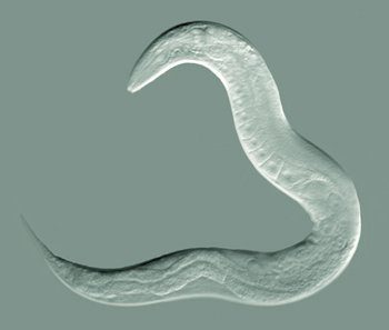
Nematodes
Definition of Nematodes:
The nematodes or roundworms constitute the phylum Nematoda (also called Nemathelminthes). They are a diverse animal phylum inhabiting a broad range of environments.
Classification of Nematodes:
1. Intestinal
A. Small Intestine
- Ascaris lumbricoides. (Round worm)
- Ankylostoma duodenale.
- Necator americanas. (Hookworm)
- Strongyloides stercoralis.
- Trichinella spiralis
B. Caecum and appendix
- Enterobias vermicularis. (Thread worm or pinworm)
- Trichuris trichiuria (Whipworm)
2. Somatic (Inside the tissues and organs)
A. Lymphatic System – Wucheria bancrofti.
B. Conjunctiva. – Loa loa. (African eye worm)
C. Subcutaneous tissue.-Loa loa.
D. Mesentery-Mansonella ozzardi.
E. Lungs – Strongyloides stercoralis
3. Larva migrans (These are the larvae that do not mature in the human host but can cause disease.
A. Visceral Larva migrans.
e.g.- Toxocara canis
(most serious)
B. Cutaneous larva migrans
e.g.- Ancylostoma caninum (less serious)
Common Characteristics of Nematodes:
- They are unsegmented worms without any appendage.
- They are elongated and cylindrical or filiform in appearance.
- Both of their ends are often pointed.
- The sites show a great variation from less than 5mm to upto 1 m.
- The body is covered with cuticle (a non-cellular, highly resistant coating).
- They possess a body cavity in which digestive and genital systems float where as excretory and nervous systems are rudimentary.
- The nematodes of man are all delicious helminths, i.e. the sexes are separate.
- The female nematode is usually larger than male.
- The male nematode typically has a coiled tail
Mode of Infection of Nematodes:
➤ By ingestion of:
- Embryonated eggs containing food and drink, as in A. lumbricoides, E. vermicularis and T. trichiura.
- Growing embryos in an intermediate host (infected cycles) as in D. medinensis.
- Encysted embryos in infected pig’s flesh – e.g. T. spiralis.
➤ By penetration of the skin: the filarifrom larvae as in A. duodenales, N. americanas and S. stercoralis.
➤ By blood-sucking insects: as in parasites belonging to the super family filarioidea.
➤ By inhalation of infected dust containing embryonated eggs: as in A. lumbricoides and E. vermicularis.
![]()