Valvular Heart Disease – Health of the children has been considered as the vital importance to all societies because children are the basic resource for the future of humankind. Nursing care of children is concerned for both the health of the children and for the illnesses that affect their growth and development. The increasing complexity of medical and nursing science has created a need for special area of child care, i.e. pediatric nursing.
Pediatric nursing is the specialized area of nursing practice concerning the care of children during wellness and illness. It includes preventive, promotive, curative and rehabilitative care of children. It emphasizes on all round development of body, mind and spirit of the growing individual. Thus, pediatric nursing involves in giving assistance, care and support to the growing and developing children to achieve their individual potential for functioning with fullest capacity.
Valvular Heart Disease
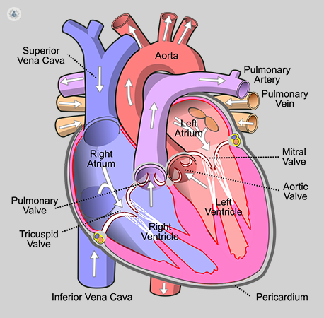
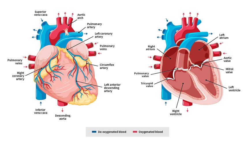
The heart has four chambers. The two upper chambers are called the left and right atrium, and the two lower chambers are called the left and right ventricle. The four valves at the exit of each chamber maintain one-way continuous flow of blood through the heart to the lungs and the rest of the body.
Valvular heart disease is any cardiovascular disease process involving one or more of the four valves of the heart (the aortic and mitral valves on the left side of heart and the pulmonic and tricuspid valves on the right side of heart).
These conditions occur largely as a consequence of ageing, but may also be the result of congenital (inborn) abnormalities or specific disease or physiologic processes including rheumatic heart disease and pregnancy.
Definition of Valvular Heart Disease:
Valvular heart disease is any cardiovascular disease process involving one or more of the four valves of the heart (the aortic and mitral valves on the left side of heart and the pulmonic and tricuspid valves on the right side of heart).
Types of Valvular Heart Disease:
Valvular stenosis (narrowing)
The stiffening of heart valves can narrow the size of the valve opening and restrict blood flow. The narrowing is called valve stenosis.
- Tricuspid valve stenosis
- Pulmonary valve stenosis
- Mitral valve stenosis
- Aortic valve stenosis
Valvular prolapse (slipping out of place)
Prolapse is a condition when the valve flaps (leaflets) slip out of place or form a bulge. This can lead to improper or uneven closure of the heart valve. As a result of the prolapsed valve, blood may leak backwards through the valve and one-way blood flow may be disrupted.
- Mitral valve prolapse
- Tricuspid, pulmonary and aortic valve prolapse
Regurgitation (leaking)
Regurgitation can happen when the valve doesn’t close properly and allows blood to flow backwards. This disruption of the one-way blood flow in the heart puts a strain on your heart, reduces its pumping efficiency and limits its ability to supply your body with oxygen-rich blood.
- Tricuspid valve regurgitation
- Pulmonary valve regurgitation
- Mitral valve regurgitation
- Aortic valve regurgitation
Causes of Valvular Heart Disease:
Congenital causes
- Congenital valvular heart disease
- Bicuspid aortic valve disease
- Marfan syndrome
Acquired causes
- Rheumatic fever
- Infective (bacterial) endocarditis
- Radiation therapy
- Age
Other causes
- Coronary artery disease
- Damage to the heart muscle from a heart attack
- Other diseases of the heart muscle (cardiomyopathy)
- Metabolic disorders such as high blood cholesterol
- Tumour in the heart
- Certain medications.
MITRAL STENOSIS (MS)
Mitral stenosis (MS) is characterized by obstruction to left ventricular inflow at the level of mitral valve due to structural abnormality of the mitral valve apparatus.
Causes of mitral stenosis:
- Rheumatic heart diseases (Principal cause)
- Calcification of mitral valve in elderly pt.
- Congenital
- Lutembacher’s syndrome (Acquired MS with ASD)
(Ref: Davidson-23rd/518)
Nice to know:
- Normal mitral orifice: 5 cm². (4-6 cm²)
Classification of MS according to mitral valve orifice:
- Minimal: mitral valve area <5 but >2.5 cm²
- Mild: mitral valve area 1.5 to 2.5 cm²
- Moderate MS: 1-1.5 cm²
- Severe MS/critical MS: < 1 cm² Symptoms at rest.
Clinical feature:
Symptom:
- Breathlessness (due to pulmonary congestion)
- Fatigue (due to low cardiac output) Oedema, ascites (right heart failure)
- Palpitation (atrial fibrillation)
- Haemoptysis (due to pulmonary congestion, pulmonary embolism)
- Cough (due to pulmonary congestion)
- Chest pain (due to pulmonary hypertension)
- Symptoms of thromboembolic complications (e.g. stroke, ischaemic limb)
Signs:
- Pulse- irregularly irregular pulse (Atrial fibrillation)
- Mitral facies (Malar flush)
- JVP-Raised JVP if developed right heart failure
- Palpation:
✓ Tapping apex beat
✓ Palpable P2 if develops pulmonary hypertension
✓ Left parasternal heave if rt. ventricular hypertrophy.
- Auscultation:
✓ Loud 1 heart sound, opening snap
✓ Mid-diastolic murmur (there is a localized low pitched, rough, rumbling, mid diastolic murmur preceded by an opening snap with a pre-systolic accentuation without any radiation best heard by the bell of the stethoscope over the mitral area patient in left lateral position breath hold after expiration)
- Signs of raised pulmonary capillary pressure:
✓ Crepitations, pulmonary oedema, effusions
Investigations of mitral stenosis:
- ECG:
✓ Left atrial hypertrophy (P mitrale)
✓ Right ventricular hypertrophy (Tall R wave in V1,V2 & deep S wave in Vs, V6)
✓ Feature of Atrial fibrillation
- Chest X-ray:
✓ Enlarged left atrium with appendage ✓ Signs of pulmonary venous congestion
- Echo:
✓Thickened immobile cusps
✓ Reduced valve area
✓ Reduced rate of diastolic filling of LV
- Doppler:
✓ Pressure gradient across mitral valve
✓ Pulmonary artery pressure
✓ Left ventricular function
- Cardiac catheterization: Assessment of coronary artery disease, pulmonary artery pressure
- MS and MR
✓ Enlarge left atrium
Treatment:
Medical treatment: [Remember =ADDA]
✓ Anti-coagulants: Warfarin
✓ Pt with systemic embolization for at least 1 yr.
✓Pt with atrial fibrillation for permanently
✓Ventricular rate control by Digoxin: 0. 25 mg/day, beta blocker or calcium channel blocker in artial fibrillation.
✓Diuretics to control pulmonary congestion & rt. heart failure.
✓ Prophylactic antibiotic against infective endocarditis (rarely recommended)
- Definitive treatment:
✓ Mitral balloon valvuloplasty: treatment of choice.
✓ Mitral valvotomy-if facilities or expertise for valvuloplasty are not available.
✓ Mitral valve replacement
- Monitoring & after care:
✓ Echocardiography
✓ ECG
✓Follow up: 1-2 yearly
(Ref by: Davidson-517-18/23rd)
Complications of Mitral Stenosis:
- Atrial fibrillation.
- Systemic thrombo-embolism eg. Stroke, gangrene of limbs, intestine.g
- Pulmonary HTN.
- Right heart failure.
- Pulmonary infarction
- Chest infection
- Tricuspid regurgitation.
- Infective endocarditis.
- Haemoptysis
- Ortner’s syndrome (Hoarseness of voice due to compression of left laryngeal nerve)
- Dysphagia (Due to oesophageal compression)
(Ref-Kumar & clark-760/8th)
Radiological Findings in Mitral Stenosis:
i. Signs of enlarged left atrium:
- Fullness of pulmonary conus leads to straightening of left heart border.
- Double contour of rt heart border.
- Upper lobe diversion.
- Widening of carina.
- Left bronchus appears horizontal.m
ii. Signs of pulmonary venous congestion
- Kerly’s B lines.
- Ground glass appearance.
iii. Calcified mitral valve may be seen in the late course of disease on a penetrated or lateral view.
(Ref-Kumar & clark-742/8)

Differences mitral stenosis from aortic regurgitation:
| Traits | Mitral stenosis | Aortic regurgitation |
| 1. Pulse | Low in volume & normal in rhythm. May be irregularly irregular (if AF). | High volume, collapsing pulse. |
| 2. Blood pressure | Normal | Wide pulse pressure. |
| 3. Face | Malar flush. | Normal face. |
| 5 1st heart sound | Loud | Normal |
| 6. Cardiac murmur | Mid-diastolic murmur at apex. | Blowing early diastolic murmur in left parasternal border at 3rd or 4th |
| 7. Pistol shot murmur over femoral artery | Absent. | Present. |
| 8. Capillary pulsation in nail beds | Absent. | Present |
Mitral Regurgitation
Mitral valve regurgitation also called mitral regurgitation, mitral insufficiency or mitral incompetence is a condition in which the heart’s mitral valve doesn’t close tightly, allowing blood to flow backward in heart.
Causes of Mitral Regurgitation:
- Rheumatic heart diseases (Principal cause-50%)
- MI
- Mitral valve prolapse
- Ischaeemia/infarction of papillary muscle
- Dilatation of the LV & mitral valve ring (Coronary artery disease, cardiomyopathy)
- Damage to valve cusps & chordae (Infective endocarditis)
Acute Causes of Mitral Regurgitation:
- Acute MI (Rupture of papillary muscle)
- Infective endocarditis
- Acute rheumatic fever
- Trauma in chest
- Cardiac surgery.
(Ref by: Davidson/23rd/519)
Management of Mitral Regurgitation.
Clinical Features of Mitral Regurgitation:
Symptoms:
- Dyspnoea (pulmonary venous congestion)
- Fatigue (low cardiac output)
- Palpitation (AF, increased stroke volume)
- Oedema, ascites (right heart failure)
Signs:
- Atrial fibrillation/ flutter
- Cardiomegaly – Displaced hyperdynamic apex beat.
- Apical pansystolic murmur + thrill
- Soft S1, apical S3
- Signs of pulmonary venous congestion (crepitations, pulmonary oedema, effusions)
- Signs of pulmonary hypertension and right heart failure
(Ref: Davidson-234/521)
Investigations mitral regurgitation:
L ECG:
- Left atrial hypertrophy (if not in AF)
- Left ventricular hypertrophy
ii. Chest X-ray:
- Enlarged left atrium
- Enlarged left ventricle
- Pulmonary venous congestion
- Pulmonary oedema (if acute)
iii. Echo:
- Dilated LA, LV
- Dynamic LV (unless myocardial dysfunction predominates)
- Structural abnormalities of mitral valve (e.g. prolapse)
iv. Doppler:
- Detects and quantifies regurgitation
v. Cardiac catheterization:
- Dilated LA, dilated LV, mitral regurgitation
- Pulmonary hypertension
- Coexisting coronary artery disease
Treatment of Mitral Regurgitation:
i. Medical Management:
- Diuretics
- Vasodilators, e.g. ACE inhibitors
- Digoxin if atrial fibrillation is present
- Anticoagulants if atrial fibrillation is present
- Antibiotic prophylaxis against infective endocarditis
ii. Surgery:
- Mitral valve replacement or repair
Complication of mitral regurgitation:
- Acute left ventricular failure
- Infective endocarditis
- Systemic embolism
- Atrial fibrillation.
Aortic Regurgitation
Aortic regurgitation (AR) is the diastolic flow of blood from the aorta into the left ventricle (LV). Regurgitation is due to incompetence of the aortic valve or any disturbance of the valvular apparatus (eg, leaflets, annulus of the aorta) resulting in the diastolic flow of blood into the left ventricular chamber.
Causes of Aortic Regurgitation:
i. Congenital:
- Bicuspid valve or disproportionate cusps
ii. Acquired:
- Rheumatic disease
- Infective endocarditis
- Trauma
- Aortic dilatation (Marfan’s syndrome, aneurysm, dissection, syphilis, ankylosing spondylitis)
(Ref by: Davidson-234/524)
Symptom & Signs of Aortic Regurgitation:
Symptoms:
i. Mild to moderate AR:
- Often asymptomatic
- Awareness of heart beat, ‘palpitations”.
ii. Severe AR:
- Breathlessness
- Angina
Signs:
i. Pulses:
- Large volume or collapsing’ pulse
- Low diastolic and increased pulse pressure
- Bounding peripheral pulses
- Capillary pulsation in nail beds – Quincke’s sign
- Femoral bruit (pistol shot) – Durozie’s sign
- Head nodding with pulse – de Musset’s sign
ii. Murmurs:
- Early diastolic murmur (best heard with the diaphragm in left 3rd/4th intercostals space with the pt. leaning
- forward and breath hold in expiration)
- Systolic murmur (increased stroke volume)
- Austin Flint murmur (soft mid diastolic)
iii. Other signs:
- Displaced, rocking apex beat (volume overload)
- Pre-systolic impulse
- 4th heart sound
- Pulmonary venous congestion (crepitation’s)
Investigations of Aortic Regurgitation
i. ECG:
- Initially normal, later LV hypertrophy and T-wave inversion
ii. Chest X-ray:
- Cardiac dilatation, maybe aortic dilatation
- Features of left heart failure
iii. Echo:
- Dilated left ventricle
- Hyperdynamic left ventricle
- Fluttering anterior mitral leaflet
- Doppler detects reflux
iv. Cardiac catheterization (may not be required)
- Dilated LV
- Aortic regurgitation
- Dilated aortic root

Treatment of Aortic Regurgitation.
- Asymptomatic patients should also be followed up annually with echocardiography
- Rx according to underlying conditions such as endocarditis or syphilis.
- Systolic blood pressure should be controlled with vasodilating drugs such as nifedipine or ACE inhibitors.
- Antibiotic prophylaxis against infective endocarditis
- Treatment of heart failure-Salt restriction, digoxin, diuretics & ACE inhibitors.
Surgical treatment:
i. Aortic valve replacement is indicated if
- Symptomatic aortic regurgitation
- Asymptomatic pt with LV ejection fraction<55% or LV end diastolic
- dimention increase to ≥ 55 mm
- Acute severe AR
ii. Sometimes combined with aortic root replacement and coronary bypass surgery may be necessary such as Marfan’s syndrome
(Ref: Davidson-524/23rd)
Read more: