Worm infestation – Health of the children has been considered as the vital importance to all societies because children are the basic resource for the future of humankind. Nursing care of children is concerned for both the health of the children and for the illnesses that affect their growth and development. The increasing complexity of medical and nursing science has created a need for special area of child care, i.e. pediatric nursing.
Pediatric nursing is the specialized area of nursing practice concerning the care of children during wellness and illness. It includes preventive, promotive, curative and rehabilitative care of children. It emphasizes on all round development of body, mind and spirit of the growing individual. Thus, pediatric nursing involves in giving assistance, care and support to the growing and developing children to achieve their individual potential for functioning with fullest capacity.
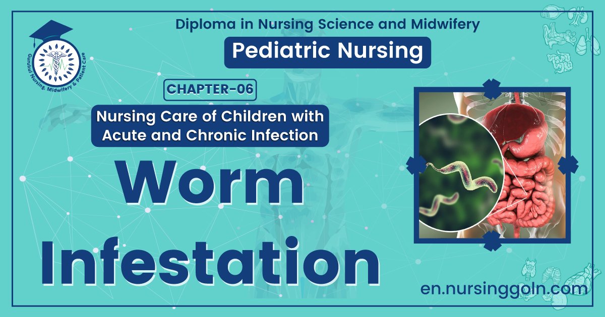
Worm infestation
Worm infestation occurs when worms live as parasitic adults in the human gastrointestinal tract. Worms that infect humans can be divided into three groups:
1. Roundworms, whipworms, hookworms
2. Tapeworms
3. Flukes
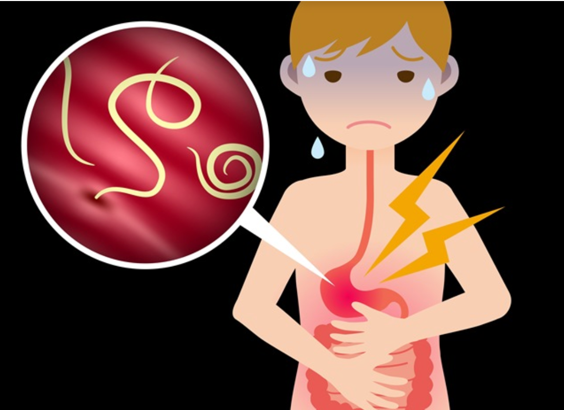
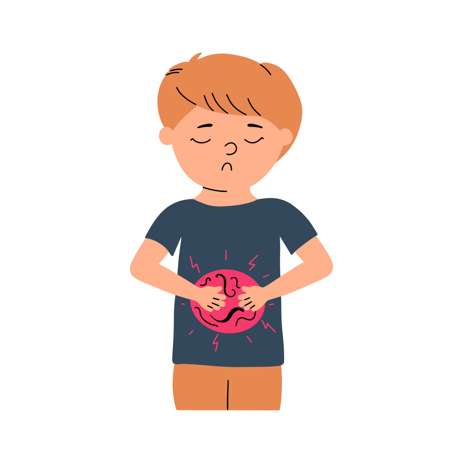
A patient with a mild infection may not show any symptom. However, some may experience itching around the anal area especially at night when the female worm deposits eggs in the perianal region. Other symptoms include poor appetite which leads to weight loss, stomach discomfort, restless sleep and inability to concentrate. A worm passed with a stool may also be discovered by the patient.
The most common route of transmission for worm infestation is the anus-to-mouth route. This is because eggs are often found under the fingernails of the infected person who has scratched the anal area. When the infected person uses the contaminated fingers to handle and ingest food, the eggs are transferred directly from the anus to the mouth.
Eggs dislodged from the perianal area into the environment can also be inhaled. Eggs may also be spread by house dust, from pets or through contact with contaminated objects such as bedding, cups, utensils and doorknobs.
Definition of Worm Infestation
Worm infestation occurs when worms live as parasitic adults in the human gastrointestinal tract. Worms that infect humans can be divided into three groups:
1. Roundworms, whipworms, hookworms
2. Tapeworms
3. Flukes
Common Worm infestations in Bangladesh/ Types of Worm Infection:
There are countless number of species in this world. These parasites, which harbor in your child’s intestine also come in various forms, shapes and sizes. Below are most common types of worm infections that trouble children:
1. Tapeworms: These are flat, ribbon-like worms that can grow up to 15-30 ft and thrive in the intestine
2. Roundworms: They resemble earthworms and can grow up to the size of 30-35 cm
3. Pinworms or thread-worms: As the name suggests, thread-worms appear to be fine white cottony threads and live in intestine and around anus of the individual
4. Hookworms: These are commonly contracted through coming in touch with the contaminated soil and later enter intestine
Symptoms of Worm Infections in Children
Apart from a stomach ache, irritability and some weight loss, worm infection can be denoted by following signs:
Tapeworm Infection
1. Nausea and vomiting
2. Loss of appetite, sometimes eating too frequently and weight loss: These usually appear in case of infection by tapeworms. Malnutrition may also be seen
3. jaundice: this is more common in tapeworm infections
Thread-worm Infection
1. Itching around anus is a common complaint in children who have thread-worm infection. Thread-worms creep out during night and lay eggs near anus which can trigger itching
2. Trouble sleeping (due to itching)
3. Painful urination: this is typical in thread-worm infestation
Roundworm Infection
1. Diarrhea, passing worms with stools: If roundworm infection persists, then mature worms may be passed out from stools
2. Fever and dry cough: These are spotted within 4-16 days after coming in contact with the roundworm eggs
Hookworm Infection
1. Coughing, wheezing: Such respiratory troubles may crop up when the hookworm larvae attack lungs
2. Anemia, fatigue: These symptoms can be seen in case of severe hookworm infection
Causes of Worm Infestation
A few common causes of worm infection include
1. Coming in contact with infected surface: The worms and their eggs are capable of surviving up to two weeks without feeding. Some of the most common places where your kid may contract worm infestation are:
soil containing worms or eggs – in playground or outdoor play touching pets or their excrement infected with worms
2. Inadequate hand washing: It’s difficult to keep young kids from putting things in their mouth. If your kid is suffering from worms, there will be itching around anus, particularly in case of pin worms. During scratching, the eggs of the worms come in contact with the skin on hand, which wherever touched tends to spread. The worse scenario is when kids put that hand back in the mouth, such as for thumb sucking or pleasure. Read more about children and
3. Improper hygiene: Unwashed bedding, undergarments, filth in the room all present breeding room for worms and their eggs. Someone who has not washed hands properly can also pose a risk for others as worm eggs can stay on fingernails which can be passed to you child through touching
4. Consuming infected food or water: It is very important to wash vegetables and fruits before consumption as worm eggs can be on them. Raw or under-cooked food also carries a risk of worm infestation. Contaminated water is again a very common source of worm infestation
Preventive Measure of Worm Infestation
1. Primary prevention
- Sanitary disposal of faeces
- Provision of safe drinking water
- Food hygiene habits
- Health education of community regarding: Use of sanitary latrines, personal hygiene, and changing behavioral patterns.
2. Secondary prevention:
- Albendazole: 400mg all ages above 2 years, Single dose
- Mebendazole: 500 mg single dose or 100 mg twice daily for 3 days.
3. Other Measure for Children:
- Inculcate the habit of frequently and thoroughly washing the hands with a good antibacterial soap
- Teach your kid to drink clean, filtered or boiled water. Practice the same at home
- Make sure your kids change their undergarments daily
- Wash their bedding, pillow covers, blanket etc. regularly
- Sterilize your toddler’s toys
- Encourage your kid to play in dry areas and not splash in muddy puddles as these horde millions of germs
- Make sure that your veggies and meat are thoroughly cooked before you serve them to your kid
- Keep your kid’s nails well-trimmed. Show them how dirt collects under long fingernails and must be kept clean
- Washing hands thoroughly before eating anything should be the rule!
- Teach potty hygiene
- Do not share towels and undergarments
- Teach your kid to shower regularly. Practice thorough cleaning of private parts
- Clean your house thoroughly and with proper disinfectants
- Allow plenty of sunshine in your kid’s room. Some worms are sensitive to light and nothing better than letting the sun do this job
Complications of Worm Infestations:
➤ Slowed growth. Loss of appetite and poor absorption of digested foods put children with ascariasis at risk of not getting enough nutrition, which can slow growth.
➤ Intestinal blockage and perforation. In heavy ascariasis infestation, a mass of worms can block a portion of your intestine. This can cause severe abdominal cramping and vomiting. The blockage can even make a hole in the intestinal wall or appendix, causing internal bleeding (hemorrhage) or appendicitis.
➤ Duct blockages. In some cases, worms may block the narrow ducts of your liver or pancreas, causing severe pain.
Tape Worms
Classification of Tape Worms/ Cestode:
It can be classified in two ways.
1. On the basis of habitat
2. On the basis of Systemic classification
Medically important cestodes are classified according to habitat. It is as follows –
According to habitat:
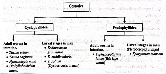
General Characters of Cestodes:
- Cestodes are segmented and tape like.
- Sizes vary from a few millimeters to several meters.
- Adult worms are found in the intestinal canal of man and animal.
- “Head” is provided with suckers (slit- like or cup-like) and sometimes with hooks which serve as organs of attachment.
- There are three regions in an adult worm: “head” (scolex), “neck” and a strobila (a body or trunk) consisting of a series of segments (proglottides).
- Sexes are not separate.
- Body cavity is absent.
- Alimentary canal is entirely absent.
- Excretory and nervous systems are present.
- Reproductive system is highly developed and complete in each segment.
Life cycle of Enterobius vermicularis:
➤Infective form: Embryonated egg containing tadpole like larva.
➤Portal of entry: Alimentary canal (Faeco-oral route).
➤ Site of location: Caecum and vermiform appendix.
➤ Types of infection:
- Person to person
- Auto infection and
- Retrograde infection
Life cycle:
- Host-Man is known host: No intermediate host is required.
Stages
Newly laid egg in perianal skin, containing tadpole like larva Completes its development within 24-36 hours in presence of O2 Infection by ingestion of these eggs In gut, egg shell dissolved by digestive juices → Larva escape in the small intestine → Develops into adolescent worm sexually matures Male fertilizes the female and dies Gravid female migrates from small intestine to caecum and colon and vermiform appendix → Remain there until egg develops → Fertilized female then wander down the rectum and works it way out of the anus at night → Deposit egg in perianal skin The cycle is repeated (Whole cycle requires 2-4 weeks ).
(Ref by- KDC/12/180)
Mode of Transmission of Enterobius Vermicularis:
Ingestion of eggs by-
- Person to person
- Auto infection and
- Retrograde infection
Clinical Conditions Caused by Enterobius vermicularis:
- In heavy infection, abdominal pain, anal pruritus, pallor and dysentery.
- Perianal pruritus and an eczematous condition around the anus and perineum.
- Nocturnal enuresis (frequency of micturition).
- Vulvitis and vulvo-vaginitis in girls.
- Sometimes penetrate into the peritoneal cavity and cause salpingitis or even, peritonitis.
- UTI among young girls.
- Sometimes inflammation of the vermiform appendix (in 2% of cases)
- Bilateral tubo-ovarian abscess.
(Ref by-KDC/12/181+ Prof. Akram/2nd/195)
Laboratory Diagnosis of Enterobiasis:
➤ Principle: Microscopic identification of pinworm eggs in the perianal region is the evidence of infection. Eggs are rarely found in the feces, and the diagnosis is made by finding eggs on perianal swabs made of Scotch tape. The tape is pressed first onto the perianal region and then onto a microscope slide, and is examined microscopically. Perianal specimens are best obtained in the morning before bathing or defecation. Three specimens should be taken on consecutive days before pinworm infection is ruled out.
➤ Specimen collection: Perianal swab.
➤ Microscopic examination:
- Coverslip preparation with saline.
- Demonstration of egg of E. vermicularis the perianal skin.
➤ Macroscopic examination:
- Adult worm may be found in the perianal region at night or during passing stool.
➤ Non specific examination:
- Occult blood test-Often positive.
- Differential count of W.B.C.-may show eosinophilia
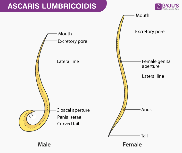
Ascaris lumbricoides
Ascaris lumbricoides is the “large roundworm” of humans, growing to a length of up to 35 cm (14 in). It is one of several species of Ascaris. An ascarid nematode of the phylum Nematoda, it is the most common parasitic worm in humans.
This organism is responsible for the disease ascariasis, a type of helminthiasis and one of the group of neglected tropical diseases. An estimated one-sixth of the human population is infected by A. lumbricoides or another roundworm.
Ascariasis is prevalent worldwide, especially in tropical and subtropical countries. It has been proposed that Ascaris lumbricoides and Ascaris suum (pig roundworm) are the same species
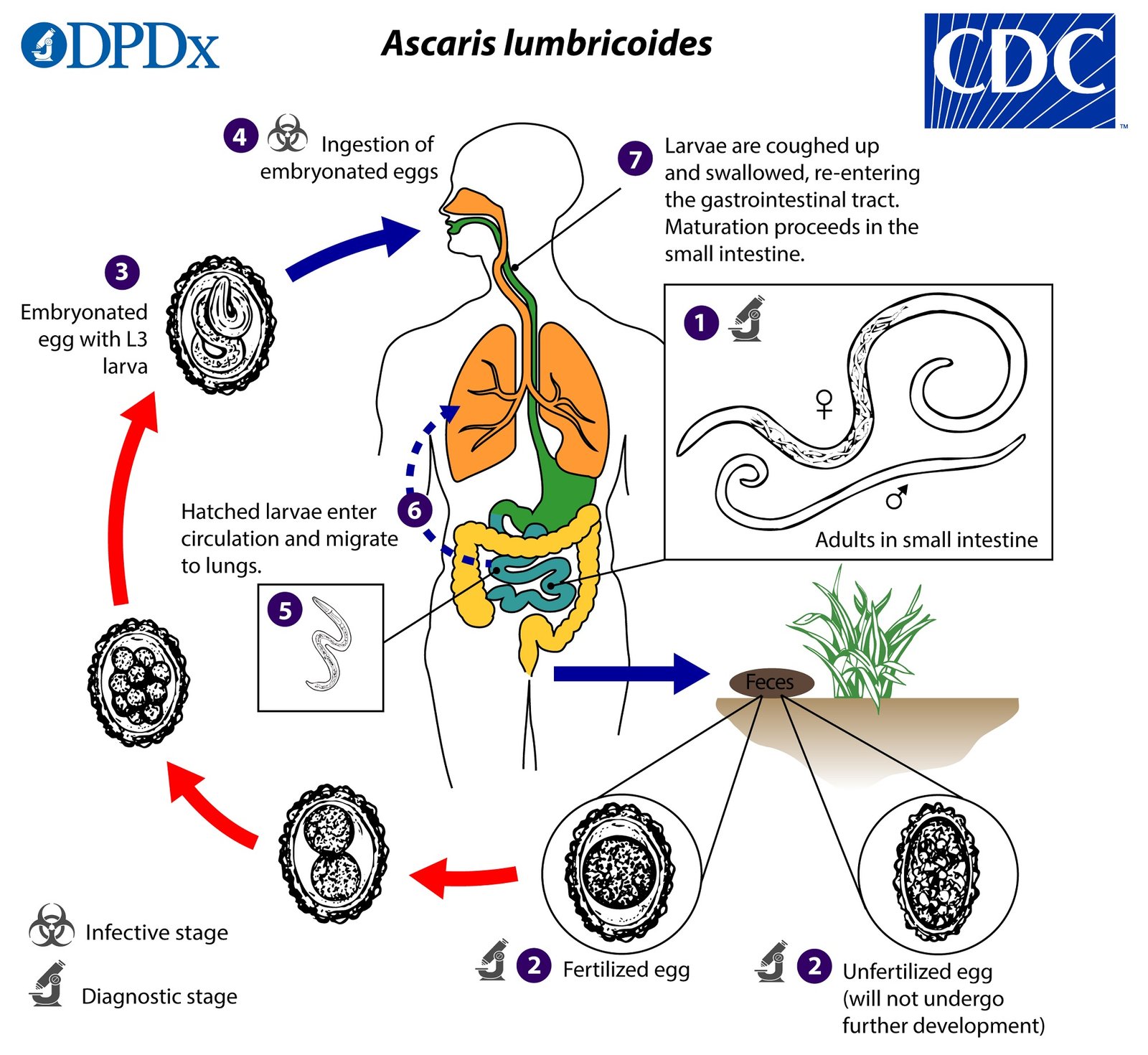
Life cycle of Ascaris lumbricoides (Round worm):
➤Infective agent: Embryonated egg.
➤Infective form: Rhabditiform larva.
➤ Mode of infection: Ingestion of contaminated foods or drinks.
➤ Portal of entry: Alimentary canal (Faeco-oral)
➤Migration: Through lungs.
➤ Site of location: Small intestine.
➤ Life cycle:
- Host – Man is the only known definitive host.
- Stages – The various stages in the life cycle are described below :
Stage 1:
Eggs in faeces –
Fertilized eggs containing the unsegmented ovum are passed with the faeces.
↓
Stage 2: Development in soil
A rhabditiform larva is developed from the unsegmented ovum within the egg shell which is infective to host. Before hatching, the larva undergoes a molting.
↓
Stage 3: Infection by ingestion and liberation of larva
When infected with food and drink or raw vegetables, the embryonated eggs pass down to duodenum and rhabditiform larvae are liberated in the upper part of small intestine.
↓
Stage 4: Migration through the lungs
The newly hatched larva burrow this way through the mucous membrane of small intestine and are carried by the portal circulation to the liver → right heart → pulmonary circulation → lung alveoli. double
↓
Stage 5: Re-entry into the stomach and the small intestine
From lung alveoli → Bronchi Trachea → larynx pharynx → swallowed stomach → supper intestine (small)
↓
Stage 6: Sexual maturity and egg liberation
Larva grow into adult worm → Sexually mature → Gravid female → Discharge egg in stool
↓
Cycle is completed and fresh cycle starts.
(Ref by- K.D.C/12/184)
Pathogenicity/Pathogenic Lesion/Clinical Features:
1. Infection of A. lumbricoides in man is known as Ascariasis.
2. The symptoms due to A. lumbricoides infection may be divided into following three groups –
- Symptoms due to adult worms.
- Symptoms due to eggs.
- Symptoms due to migrating larvae.
They are discussed below:
Symptoms due to adult worms:
➤ Asymptomatic or mild symptoms.
➤ GI tract discomfort: Anorexia, nausea, vomiting.
➤ Spoliative action: Protein-energy malnutrition (PEM) with vitamin A deficiency (Night blindness).
➤ Toxic action: The body fluid of Ascaris when absorbed is toxic and may give rise to typhoid like fever.
➤ Allergic manifestations: Hypersensitivity reaction –> Ascaris encephalopathy.
➤ Mechanical Effect: Intestinal obstruction, intussusception.
➤ Due to migration (Ectopic Ascariasis):
- Suffocation – if ascariasis may accidentally enters into respiratory passage blocks rima glottidis or enters into bronchules.
- Ascaris may come out through mouth and nose.
- Wondering ascariasis: Appendicitis, obstructive jaundice, acute haemorrhagic pancreatitis, liver abscess etc.
Symptoms due to eggs:
➤ Eggs do not cause any lesion in the intestine.
➤ Ova in the biliary canaliculi and liver may produce inflammatory changes.
Symptoms due to migrating larvae:
➤ Bronchitis and pneumonitis.
➤ Loeffler’s syndrome.
Characteristics of Eggs of Ascaris:
Characteristics of fertilized egg:
- Round or oval in shape.
- Bile-stained and brownish (golden brown) in colors.
- Surrounded by thick translucent shell with an outer albuminous coat.
- Contains a very large conspicuous, unsegmented ovum.
- Floats in saturated solution of common salt.
Characteristics of unfertilized egg:
- Narrower, longer and more elliptical.
- Brownish in colour (bile-stained).
- Has a thinner shell with an irregular coating of albumin.
- Contains a small atrophied ovum with a mass of disorganized, highly refractile granules of various sizes.
- Does not float in salt solution.
(Ref by-KDC/12th /183)
Laboratory diagnosis of A. lumbricoides:
- Adult worm: Identification of the adult worms passed through the rectum or from some other body orifice. Abdominal ultrasound may reveals worms in the gastrointestinal or biliary tracts.
- Eggs: In the stool, vomitus, sputum, or small bowel aspirate.
- Larva: Diagnosis during the stage of larval migration is difficult, although occasionally larvae may be found in the sputum or gastric contents.
- Eosinophil count: An increased number of circulating eosinophil’s may be found during the stage of larval migration.
Complications of Ascaris lumbricoides infection:
Medical condition:
- Ascaris pneumonia (Loeffler’s syndrome).
- Lesions in the various organs like brain, heart and kidney due to larvae in systemic circulation.
- Protein-energy malnutrition.
- Night blindness.
- Typhoid like fever due to absorption of toxic body fluid of ascaris lumbricoides.
- Allergic manifestations.
- Inflammatory changes in the liver and biliary canaliculi due to involvement of the ova.
Surgical condition:
- Intestinal obstruction specially in children due to relative small size of the intestinal lumen
- Intussusception
- Ulceration of alimentary canal
- Suffocation
- Appendicitis, obstructive jaundice, acute haemorrhagic pancreatitis due to wondering ascariasis.
Control and prevention:
1. Primary prevention
- Sanitary disposal of faeces
- Provision of safe drinking water
- Food hygiene habits
- Health education of community regarding: Use of sanitary latrines, personal hygiène, and changing behavioural patterns.
2. Secondary prevention:
- Albendazole: 400mg all ages above 2 years, Single dose
- Mebendazole: 500 mg single dose or 100 mg twice daily for 3 days.
- Pyrantel pamoate: 10mg/kg body weight (maximum 1 g) daily for 3 days.
- Levamisole: 50-150 mg (3 mg/kg body weight) single dose
- Mass treatment: Periodic deworming at interval of 2-3 months may be undertaken in the highly prevalent area. It will reduce worm load of the community.
Trichomonas Vaginalis
Trichomonas vaginalis causes Trichomoniasis.
Habitat:
- In the female genital tract (Primarily vagina).
- In the urinary tract of female and male (Primarily prostate).
Morphology:
- Pear shaped.
- Only trophozoite form and no cystic stage.
- A central nucleus and four anterior flagella.
- It has an undulating membrane that extends about two-thirds of its length.
Mode of transmission:
- Sexual intercourse.
Pathogenicity:
- In women: A watery, foul-smelling, greenish vaginal discharge accompanied by itching and burning sensation.
- In men: Infection is asymptomatic. About 10% of man have urethritis.
Laboratory Diagnosis:
Principle:
Demonstration of the parasite by microscopic examination and immunofluroescence are used for laboratory diagnosis. Detection of parasite by culture is helpful in asymptomatic cases. DNA based tests (PCR) are also available.
[Ref-Prof. Akram/2/76]
Specimen collection:
- In female, high vaginal swab.
- In male, morning urethral discharge.
Microscopic examination:
- Slide prepared with normal saline: Findings – Trophozoite can be found.
- Wet mount preparation of vaginal or prostatic secretion: Finding – the pear shaped trophozoite with a typical jerky motion.
Serological test: No role.