
The human skeletal system is a marvel of engineering. Among its many components, joints are crucial structures that provide mobility and flexibility. These articulations, where bones meet, vary in structure and function. One of the most common and versatile types of joints in the human body is the synovial joint. In this article, we will delve into the anatomy of selected synovial joints and understand their unique features and functions.
Anatomy of Selected Synovial Joints
Introduction to Synovial Joints
Synovial joints, also known as diarthrosis joints, are the most mobile type of joint in the human body. They possess a unique structure that allows for a wide range of motion. These joints are encapsulated within a fibrous joint capsule filled with synovial fluid, which lubricates the joint to reduce friction between the articulating bones.
Components of Synovial Joints:
- Joint Capsule: Made up of an outer fibrous layer and an inner synovial membrane. The fibrous layer provides strength, while the synovial membrane secretes synovial fluid.
- Synovial Fluid: A viscous fluid that lubricates the joint and nourishes the articular cartilage.
- Articular Cartilage: A smooth, white tissue that covers the ends of bones where they come into contact. Its primary function is to reduce friction at the joint.
- Ligaments: These are fibrous bands of tissue that connect bone to bone, providing stability to the joint.
- Tendons: Connect muscle to bone, aiding in joint movement.
- Menisci and Bursae: Some synovial joints contain these additional structures to reduce friction and cushion the joint.

Selected Synovial Joints and Their Anatomy
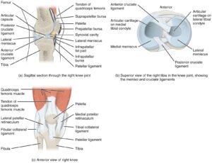
1. Knee Joint
The knee is one of the largest and most complex joints in the body. It is a hinge joint that primarily allows for flexion and extension, with a slight capacity for medial and lateral rotation.
- Bones Involved: Femur (thigh bone), Tibia (shin bone), and Patella (knee cap).
- Key Structures:
- Menisci: These are crescent-shaped fibrocartilaginous structures that cushion the joint and help distribute the load across the joint.
- Anterior Cruciate Ligament (ACL) and Posterior Cruciate Ligament (PCL): These ligaments provide anterior and posterior stability to the knee.
- Medial and Lateral Collateral Ligaments: Stabilize the knee against sideward movement.
2. Shoulder Joint (Glenohumeral Joint)
The shoulder joint is a ball-and-socket joint that offers a wide range of motion, making it one of the most mobile joints in the body.
- Bones Involved: Humerus (upper arm bone) and Scapula (shoulder blade).
- Key Structures:
- Rotator Cuff: A group of four tendons and muscles that surround the shoulder joint, providing stability and allowing rotational movement.
- Glenoid Labrum: A fibrocartilaginous rim around the glenoid cavity that deepens the socket and increases joint stability.
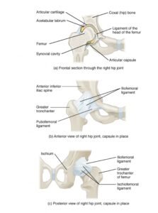
3. Hip Joint
Another ball-and-socket joint, the hip joint, is crucial for weight-bearing and locomotion.
- Bones Involved: Pelvic bone (specifically the acetabulum) and Femur (head).
- Key Structures:
- Acetabular Labrum: Similar to the glenoid labrum in the shoulder, this deepens the socket and stabilizes the joint.
- Ligament of the head of the femur (Ligamentum teres): Provides minimal structural integrity but contains a vital artery supplying the head of the femur.
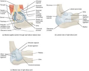
4. Elbow Joint
This hinge joint allows for the flexion and extension of the forearm.
- Bones Involved: Humerus, Radius, and Ulna.
- Key Structures:
- Radial and Ulnar Collateral Ligaments: These provide lateral and medial stability to the elbow.
- Olecranon Process: A bony prominence of the ulna that fits into the olecranon fossa of the humerus during extension.
5. Wrist Joint (Radiocarpal Joint)
The wrist joint is a complex condyloid joint, allowing for flexion, extension, abduction, and adduction.
- Bones Involved: Radius and the first row of carpal bones (Scaphoid, Lunate, and Triquetrum).
- Key Structures:
- Scapholunate and Lunotriquetral Ligaments: Maintain the alignment of the carpal bones.
- Triangular Fibrocartilage Complex (TFCC): Cushions the ulnar side of the wrist and provides a smooth surface for joint movement.


Synovial joints play a pivotal role in our daily activities, from walking and writing to dancing and lifting. Understanding their anatomy and function not only provides insight into our body’s remarkable capabilities but also emphasizes the importance of joint health and injury prevention.
While the selected joints discussed above represent only a portion of the synovial joints present in our body, they underscore the diversity and specialization of these structures. As research progresses, our knowledge of these intricate joints and their associated conditions will undoubtedly expand, paving the way for better therapeutic strategies and treatments.