The appendicular skeleton consists of the bones of the limbs and the pectoral and pelvic girdles. The pectoral girdle connects the upper limb to the body trunk, while the pelvic girdle, which is the focus of this article, secures the lower limb. The pelvis is a basin-shaped structure that supports the spine and protects the abdominal and pelvic organs. The pelvic girdle and pelvis are complex, vital structures responsible for weight-bearing, protection, and a myriad of movements and functions.
The Pelvic Girdle and Pelvis

Anatomy of the Pelvic Girdle
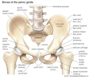
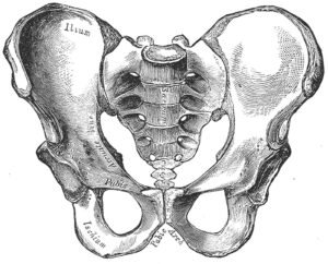
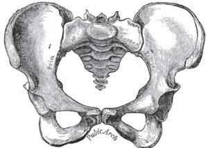
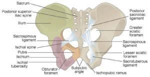
The pelvic girdle consists of two hip bones, each of which is formed by the fusion of three bones: the ilium, ischium, and pubis. These bones are separate in childhood but fuse in early adulthood.
- Ilium: The largest of the three bones, the ilium forms the uppermost part of the hip bone. Its most prominent feature is the iliac crest, which begins at the anterior superior iliac spine and ends at the posterior superior iliac spine.
- Ischium: This bone forms the lower and posterior part of the hip bone. The most familiar feature is the ischial tuberosity, which is what we sit on.
- Pubis: Located anteriorly, the pubis forms the front part of the hip bone. The two pubic bones of each hip are joined together at the midline by a cartilaginous joint called the pubic symphysis.
Together, these fused bones form a socket known as the acetabulum, where the head of the femur articulates, facilitating movement at the hip joint.
Anatomy of the Pelvis
The pelvis is made up of the two hip bones (which are part of the pelvic girdle), the sacrum, and the coccyx. Based on the position and orientation, the pelvis can be divided into two parts:
- Greater (False) Pelvis: Found above the pelvic brim, it’s bounded by the flare of the iliac bones and supports the intestines (mainly ileum and sigmoid colon).
- Lesser (True) Pelvis: Below the pelvic brim, it’s surrounded by the bone and consists of the pelvic cavity, continuing downwards to the pelvic outlet. This is the part through which a baby passes during childbirth.
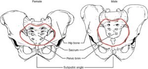
Distinguishing Features between Male and Female Pelves
Given its role in childbirth, the female pelvis is structurally different from the male pelvis.
- Pelvic Inlet: In females, it’s wider and more oval, allowing for childbirth. In males, it’s heart-shaped and narrower.
- Pelvic Outlet: Wider in females to accommodate childbirth.
- Subpubic Angle: This angle is more than 90° in females and less than 90° in males. This makes the female pubic arch wider than the male’s.
- Coccyx: The male coccyx is relatively more vertical, while the female coccyx is more movable and projects backwards.
- Acetabulum: It’s larger in males compared to females.
Functions of the Pelvic Girdle and Pelvis
- Support: The pelvis provides support to the upper body’s weight, transferring it to the lower limbs during standing, walking, or running.
- Protection: It encloses and protects the pelvic and certain abdominal organs, including the bladder, reproductive organs, and rectum.
- Locomotion: With the articulation of the femur at the acetabulum, it aids in walking, running, and various lower limb movements.
- Childbirth: The female pelvis is specially adapted to allow the passage of the fetus during childbirth.
Clinical Significance
Understanding the anatomy of the pelvic girdle and pelvis is essential for medical professionals. Some areas of clinical importance include:
- Fractures: Falls, especially in elderly individuals with osteoporosis, can lead to fractures of the pelvis. In younger individuals, high-energy traumas, like car accidents, can result in pelvic fractures.
- Osteoarthritis: Wear and tear of the hip joint can cause osteoarthritis, leading to pain and reduced mobility.
- Sacroiliac Joint Dysfunction: This joint, formed between the sacrum and ilium, can cause lower back and leg pain if it becomes inflamed or if there’s too much or too little movement.
- Childbirth Complications: A narrow pelvis can result in difficulties during childbirth, sometimes necessitating a cesarean section.
The pelvic girdle and pelvis are integral components of the appendicular skeleton with vital functions in support, movement, and protection. Their complex anatomy, especially the variations between males and females, makes them central to studies in anatomy, anthropology, and medicine. An understanding of their structure and function aids in the diagnosis and treatment of various medical conditions and in appreciating the evolutionary adaptions that have facilitated bipedal locomotion and childbirth in humans.